INTERVENTIONAL FLASHLIGHT

DOI: 10.4244/EIJ-D-16-00851

Stenting complex aortic coarctation: simulation in a 3D printed model

Francesca Romana Pluchinotta*, MD; Luca Giugno, MD; Mario Carminati, MD

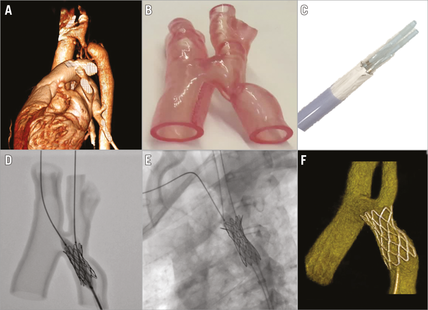
A 23-year-old lady with aortic coarctation was referred for percutaneous treatment. Cardiac CT demonstrated severe coarctation distal to the origin of the left carotid artery (LCA) with hypoplasia of the left subclavian artery (Panel A). The planned procedure was to implant a covered stent to reduce the risk of aortic dissection or rupture. However, using a covered stent, the risk of occluding the origin of the LCA had to be considered. An innovative approach was planned and tested in a 3D model printed in HeartPrint® Flex material (Materialise, Leuven, Belgium) (Panel B). A single covered CP Stent™ 39 mm (NuMED, Hopkinton, NY, USA) was mounted on, delivered over, and expanded on two separate, adjacent balloons (Cristal Balloon 10x40 mm; Balt, Montmorency, France) (Panel C). The two balloon wires were advanced into the right subclavian and the LCA (Panel D). The distal portion of the stent was expanded first to dilate the stenotic point without compromising the flow into the LCA. Then, by withdrawing the sheath, the remaining part of the stent was expanded. Later the patient underwent catheterisation. Based on aortographic diameters, the same stent and balloon used in the simulation were chosen. Stent position was easy to work out, as it had already been tested in the 3D model (Panel E). After successful stent delivery, 3D rotational angiography confirmed adequate stent expansion (Panel F). These images show the first case of a single CP covered stent mounted on two adjacent balloons in a patient with severe coarctation very close to the LCA. The 3D model was helpful in planning the intervention, and in determining the stent length and its optimal position.

Conflict of interest statement

The authors have no conflicts of interest to declare.

Volume 13 Number 4
Jul 20, 2017
Volume 13 Number 4
View full issue


Key metrics

Suggested by Cory

EXPERT REVIEW

10.4244/EIJV11I6A133 Oct 20, 2015
The effectiveness of stenting of coarctation of the aorta: a systematic review
Hartman E et al
free

IMAGE IN CARDIOLOGY

10.4244/EIJ-D-16-00570 Dec 20, 2016
Percutaneous repair of severe coarctation of the aorta
Biswas S et al
free

Clinical research

10.4244/EIJ-D-18-00923 Mar 20, 2020
Percutaneous reintervention on aortic coarctation stenting
Pan M et al
free

Clinical research

10.4244/EIJ-D-19-00157 Feb 5, 2021
Early and midterm outcomes of bare metal stenting in small children with recurrent aortic coarctation
van Kalsbeek R et al
free
Trending articles
318.3

State-of-the-Art Review

10.4244/EIJ-D-21-00695 Nov 19, 2021
Transcatheter treatment for tricuspid valve disease
Praz F et al
free
116.85

State-of-the-Art

10.4244/EIJ-D-24-00066 Apr 21, 2025
Management of complications after valvular interventions
Bansal A et al
free
108.3

Viewpoint

10.4244/EIJ-E-22-00007 May 15, 2022
TAVI at 20: how a crazy idea led to a clinical revolution
Eltchaninoff H et al
free
91.6

Image – Interventional flashlight

10.4244/EIJ-D-22-00344 Aug 5, 2022
First dedicated transcatheter leaflet splitting device: the ShortCut device
Tchétché D et al
free
72.15

State-of-the-art

10.4244/EIJ-D-22-00627 Feb 6, 2023
Left atrial appendage occlusion
Holmes D et al
free
68.9

State-of-the-Art

10.4244/EIJ-D-24-00992 Sep 15, 2025
Antithrombotic therapy in complex percutaneous coronary intervention
Castiello D et al
free
49.55

CLINICAL RESEARCH

10.4244/EIJ-D-17-00962 Apr 6, 2018
A new optical coherence tomography-based calcium scoring system to predict stent underexpansion
Fujino A et al
free
42.95

State-of-the-Art Review

10.4244/EIJ-D-21-00145 Sep 20, 2021
Robotics, imaging, and artificial intelligence in the catheterisation laboratory
Beyar R et al
free
X

PCR
Impact factor: 9.5
2024 Journal Citation Reports®
Science Edition (Clarivate Analytics, 2025)
Online ISSN 1969-6213 - Print ISSN 1774-024X
© 2005-2025 Europa Group - All rights reserved