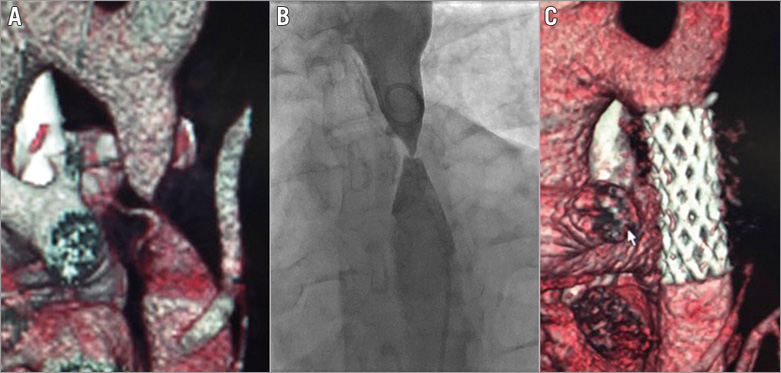

A 21-year-old asymptomatic male was referred with severe hypertension. Blood pressure was 190/100 mmHg in both arms. Examination revealed absent femoral and pedal pulses. Electrocardiography showed sinus rhythm with left ventricular hypertrophy. Echocardiography demonstrated moderate left ventricular hypertrophy with a trileaflet aortic valve. Computed tomographic aortography (CTA) demonstrated severe discrete coarctation with a diameter of <1 mm at the aortic isthmus (Panel A), and invasive aortography (via dual injections) demonstrated complete aortic occlusion (Panel B).
Transcatheter intervention was performed via both radial and femoral arterial access (Moving image 1). The occlusion was crossed from above with an exchange length 0.035-inch straight wire (Terumo Corp., Tokyo, Japan), which was snared in the descending aorta (EN Snare®, 12-20 mm; Merit Medical, Galway, Ireland) and externalised to allow delivery of a 14 Fr Mullins sheath (Medtronic, Dublin, Ireland). An 8-zig 45 mm covered Cheatham-Platinum stent (NuMED, Hopkinton, NY, USA) was deployed on a 16 mm balloon without predilation. The invasive peak-peak gradient dropped from 60 to 0 mmHg. At one year, the patient remains well and is normotensive on one antihypertensive agent. Repeat CTA confirmed no aortic wall injury (Panel C).
Conflict of interest statement
The authors have no conflicts of interest to declare.
Supplementary data
Moving image 1. Video demonstrating percutaneous intervention on the coarctation of the aorta.
Supplementary data
To read the full content of this article, please download the PDF.
Video demonstrating percutaneous intervention on the coarctation of the aorta.

