Rotational atherectomy (RA) was invented by David Auth more than 30 years ago with the aim of improving the results of plain old balloon angioplasty (POBA) by plaque removal or “debulking” instead of disrupting and compressing coronary atheroma. The mechanism of RA is “differential cutting”, selectively ablating non-compliant atheromatous tissue (fibrotic or calcified), while elastic vascular tissue is spared by the Rotablator™ (Boston Scientific, Marlborough, MA, USA) burr. Since the first RA application in human coronary arteries in 1988, RA has become the embodiment of the myth of Sisyphus, who was punished by being made to push a huge stone up a hill only for it to roll down every time it neared the top, repeating this process for eternity.
The first period of pushing the “Rotastone” uphill (Figure 1) involved testing the value of RA in the treatment of coronary lesions with large burrs, alone or followed by low-pressure balloon dilatation, as opposed to POBA. Stents were not available at this time. Although RA achieved a high level of procedural success with complication rates similar to those for POBA, this initial benefit diminished in the medium term as the approximately 40% restenosis rate was similar for both treatments1,2,3. Consequently, the stone rolled back down the slope.
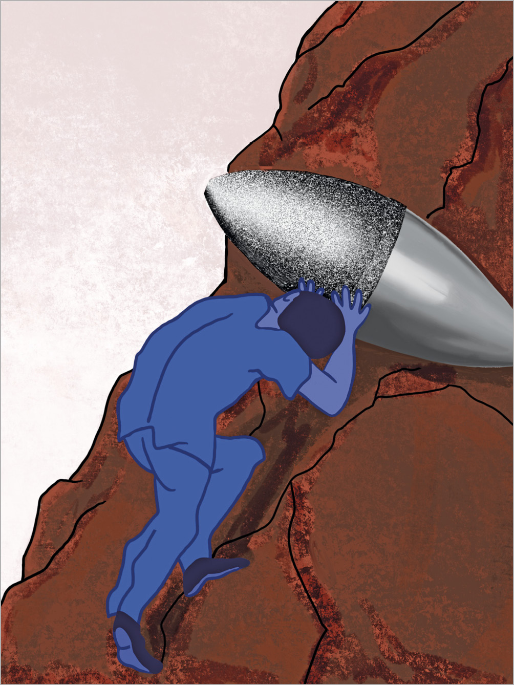
Figure 1. Sisyphus pushing the “Rotastone” uphill.
The advent of metal coronary stents in the early 1990s raised the expectation that plaque debulking before stenting would facilitate stent expansion and provide better long-term results than POBA. The stone was thus pushed up again. Randomised trials showed better initial results with RA but no benefits in terms of major adverse coronary events (MACE) and target lesion revascularisation (TLR) at midterm follow-up, so the stone rolled down again4.
The next period was that of the treatment of bare metal in-stent restenosis. Two randomised trials comparing RA with large burrs to retrieve intra-stent neointimal proliferation as opposed to POBA mirrored the results observed in native coronary arteries, with a small benefit in favour of RA that was not large enough to warrant a change5,6. The advent of coronary brachytherapy and, later on, of drug-coated balloons triggered the descent of the stone once again.
This initial aggressive strategy of debulking atheroma by large burrs for a broad spectrum of coronary lesions with a large plaque burden gradually led to a more conservative approach using smaller burrs (burr/artery ratio <0.7) to reduce calcified plaque volume, to fracture calcium nodules and to facilitate lesion dilatation by balloon. This new strategy, called “plaque modification” instead of “debulking”, was encouraged by two randomised trials, STRATAS7 and CARAT8, showing that procedural success, lumen enlargement and target vessel revascularisation (TVR) rates were similar for both strategies, while serious angiographic complications were much less common with small burrs. The emergence of drug-eluting stents (DES) in the early 2000s pushed the stone uphill again. Several case series reported intermediate and long-term outcomes after DES with adjunctive RA in complex calcified coronary lesions. Most of these studies reported remarkable MACE rates below 15% and TLR rates <10% within one to two years9,10. The ROTAXUS trial11 was the first study to compare RA to POBA before TAXUS™ (Boston Scientific) DES implantation in calcified coronary lesions. RA was more effective at improving lumen dimensions early on, but this was offset by a subsequent increase in proliferative response. Late lumen loss, the primary endpoint, was higher in the RA group (0.44±0.58 vs 0.31±0.52 mm; p=0.04). Rates of TLR and other cardiovascular events did not differ significantly between the groups. However, the ROTAXUS trial had important limitations: >50% of the patients had only mild to moderate calcification and therefore did not obtain any advantage from RA, and 12.5% crossed over from balloon to RA because of failure of balloon or stent delivery or suboptimal balloon expansion. Nevertheless, the conclusion was that RA did not increase the efficacy of DES in calcified lesions, and yet again the Sisyphus stone went rolling back down.
Recently, the PREPARE-CALC trial12 compared RA to modified balloons (MB: cutting or scoring) prior to latest-generation DES in calcified lesions. In contrast to previous trials, only severely calcified lesions by angiographic criteria were included. In addition, the current rotablation technique was employed (small burrs, lower rotational speeds, and high-pressure balloon dilatation before stenting). As in ROTAXUS, a higher success rate was achieved with RA (98% vs 81%), largely due to delivery failure of the bulky MB. At nine months, in-stent late lumen loss was much lower than for ROTAXUS but it did not differ between the groups (0.22±0.40 mm for RA vs 0.16±0.39 mm for MB). MACE rates were also much lower than previously reported and similar between groups. The limitations of this study were a 16% crossover from MB to RA because of uncrossable or undilatable lesions. From this trial it was clear that the effect on long-term outcomes does not rely on RA, but rather much more on newer-generation DES, provided that a suitable stent expansion is obtained.
In fact, previous randomised trials further emphasised that RA before stenting has no positive effect on late loss, restenosis or MACE issues compared to balloons. But is this really the purpose of RA? The answer is clearly no. The real purpose of RA today is to facilitate DES deployment in complex calcified lesions where we may anticipate or suspect problems with balloon dilatation or stent delivery and expansion. Consequently, there is no room for randomised trials of patients with severely calcified lesions undergoing RA versus other techniques, mainly because it is impossible to design a trial when the operators know in advance that treatment with balloon angioplasty or MB will either fail or provide only a modest but incomplete dilatation. No benefit should be expected from RA when calcification is mild or moderate. In practice, the severity of calcification is graded by qualitative assessment of angiography, and severe calcification is defined as “radio-opacities noted without cardiac motion before contrast injection”12. Angiography is less sensitive than intravascular ultrasound (IVUS), but severe calcification predicts a large calcium arch on IVUS. Even in severely calcified lesions, the benefit of RA is attenuated if the calcified lesion is not a ring of calcium covering almost three quarters of the IVUS image13. Intravascular imaging with IVUS or optical coherence tomography (OCT) further permits discrimination of superficial or deep calcium and delineation of the calcium arch. However, it could be difficult to perform these techniques before intervention since the imaging probe will not cross the lesion in most cases.
We have probably been pushing the stone up the slope all this time by the wrong path, the one that seeks clinical or angiographic benefits from RA in the long run. It is therefore worth considering a change of strategy. The article published in this issue of EuroIntervention by the Euro4C group14 represents the logical and foreseeable route for pushing the “Rotastone” along a path that allows us to put an end to this technique’s Sisyphus myth once and for all.
The origins of Euro4C date back to 2011 with the creation of the EuRota club by experts in the treatment of severely calcified lesions by RA, who published the first European consensus on the rotablation technique in this journal15. This group recently broadened its field of interest and was renamed Euro4C for its focus on Cardiac Care of Calcified and Complex patients.
The Euro4C registry reports clinical outcomes of RA in 996 patients with severely calcified lesions from 19 centres in eight European countries from October 2016 to July 2018. This registry allows the assessment of the current practice and use of RA in experienced European centres. As expected, patients were elderly (74.5 years) and at high risk considering that 25% had significant left main stenosis, 28% had three-vessel disease, 29% had chronic total occlusion, 43% were diabetic, 34% had renal failure, and 42% showed a left ventricular ejection fraction (LVEF) <50%. In such a complex population, RA was performed by the radial approach using a 6 Fr guiding catheter in 75% of the cases, a remarkable difference with respect to previous trials in which the femoral approach with 7 Fr guiding catheters predominated. A small 1.5 mm burr was the most common device used, aiming at plaque modification. Clinical success was very high (92%), with <5% in-hospital MACE and a low flow/no flow of 1.2%, reflecting how a meticulous application of the current technique according to the consensus document15 can translate into extremely good results.
At one-year follow-up, MACE was 13.2%, driven mainly by cardiac death (5.7%) and myocardial infarction (4.7%), all related to the patients’ high-risk profile, with left main disease, renal failure, acute coronary syndrome (ACS) on presentation, and low LVEF as independent predictive factors.
Although RA is not a complex technique, its regular use is required to achieve optimal results in patients with heavily calcified lesions such as those reported in this issue. Consequently, the best strategy is to have a low threshold for planned RA by using the angiographic definition of severe calcification. The adoption of a provisional strategy involving the performance of RA only in cases of undilatable lesions or when devices do not cross would reduce the number of RA cases, compromising expertise. Although complication rates remain low even in less experienced centres, high-volume centres (>30 cases/year) show the best results16. Moreover, a provisional RA strategy provides less favourable in-hospital outcomes and an increase in procedural time, fluoroscopy and resources17. In conclusion, planned or upfront RA can be carried out safely by the radial approach with excellent results following experts’ recommendations and should be adopted as the default strategy for patients with severely calcified lesions by angiographic criteria.
Conflict of interest statement
The authors have no conflicts of interest to declare.
Supplementary data
To read the full content of this article, please download the PDF.