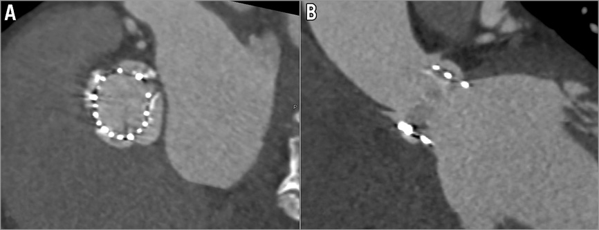
Quadricuspid aortic valve (QAV) is a rare congenital malformation and is often associated with other congenital heart anomalies1. QAV is frequently associated with regurgitation but stenosis is very unusual2-4. An 80-year-old male patient presented with multi-organ failure. Echocardiography showed a severe left ventricular systolic dysfunction with moderate mitral regurgitation and severe aortic stenosis. To save the patient, we performed balloon aortovalvuloplasty as a bridge therapy to definite aortic valve replacement, in order to minimise the procedure. Thereafter, he fully recovered from the multi-organ failure. For pre-TAVI assessment, computed tomographic angiography (CTA) (Figure 1A, Figure 1B) and transoesophageal echocardiography (Figure 1C, Moving image 1) were performed. We found the stenosed QAV without other congenital heart anomalies. The patient underwent implantation of a 26 mm Edwards SAPIEN XT valve (Edwards Lifesciences, Irvine, CA, USA) via a transfemoral approach with the guidance of Heart Navigator System (Heart Navigator 1.0.0; Philips Healthcare, Best, The Netherlands) for optimal valve positioning (Moving image 2). The CTA confirmed the proper positioning of the valve (Online Figure 1).

Figure 1. Multi-planar reconstructed computed tomographic angiographic images (A, B) and midoesophageal short-axis view (C) demonstrating quadricuspid aortic valve with thickened leaflet margin, 4 separate leaflets, and moderate calcifications.
Conflict of interest statement
The authors have no conflicts of interest to declare.
Online data supplement
Moving image 1. Midoesophageal short-axis view of transoesophageal echocardiography showed a quadricuspid aortic valve with four equal-sized cusps, a central coaptation defect, central aortic regurgitation and limited motion of aortic valve leaflets.
Moving image 2. Transcatheter aortic valve implantation was performed by guidance of Heart Navigator System, transoesophageal echocardiogram and aortogram for optimal valve positioning.

Online Figure 1. Postoperative follow-up computed tomographic angiography to ensure proper positioning of the valve: short-axis view (A) and sagittal view (B).

