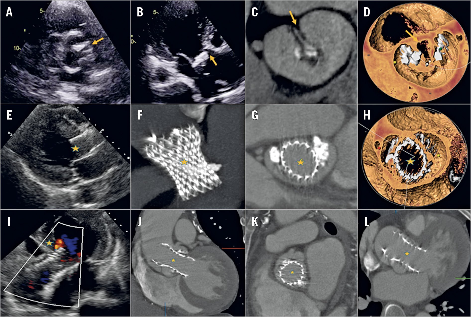
Figure 1. Illustration of echocardiogram and CT images. Transthoracic echocardiogram parasternal short-axis (A, Moving image 1) and three-chamber views (B, Moving image 2) showing calcific unicuspid, unicommissural aortic valve with an eccentric opening (arrow). Computed tomography maximum intensity projection (MIP, C) and volume-rendered (VR, D) images showing an unicuspid unicommissural valve and absence of raphae. E) Post TAVI, TTE parasternal long-axis images showing the prosthetic valve in situ. High-resolution CT MIP and VR images showing oval configuration and good expansion of the prosthetic valve (F-H, star, Moving image 3). Post TAVI, TTE five-chamber image showing the prosthetic valve in situ and showing mild paravalvular leak (I, star, Moving image 4). CT image - coronal, sagittal and axial views showing the prosthetic valve in situ (J-L, star).
A 55-year-old male presented with acute left ventricular failure. His transthoracic echocardiogram (TTE) showed a calcific, unicuspid, unicommissural aortic valve with severe stenosis and moderate regurgitation, severe biventricular dysfunction and a dilated ascending aorta (Figure 1, Moving image 1-Moving image 4). After medical stabilisation, he was advised to have surgical aortic valve and aortic root replacement. He was refused surgery in view of severe biventricular dysfunction. He underwent successful transcatheter aortic valve implantation (TAVI) with a 29 mm CoreValve® Evolut™ R (Medtronic, Minneapolis, MN, USA). His post-procedure period was uneventful.
An unicuspid aortic valve is a rare anomaly and the unicommissural variant presents with aortic stenosis in adulthood. Similar to a bicuspid aortic valve, it is associated with severe valve calcification, eccentric opening and aortopathy. Surgical aortic valve replacement along with aortic root replacement remains the appropriate treatment for these patients1. However, TAVI may be considered in patients with high surgical risk and suitable anatomy. The presence of aortopathy, a horizontal aortic root, and an eccentric valve opening makes valve crossing and device delivery challenging. In addition, unicommissural anatomy may result in non-circular expansion of the stent frame at the level of the annulus. Hence, a self-expanding supra-annular valve may offer better haemodynamic performance.
Conflict of interest statement
The authors have no conflicts of interest to declare.
Supplementary data
To read the full content of this article, please download the PDF.
Moving image 1. Parasternal short-axis view of the native valve.
Moving image 2. Three-chamber view showing the native valve.
Moving image 3. Post-TAVI CT of the prosthetic valve.
Moving image 4. Five-chamber image of the prosthetic valve.