A 76-year-old female with atrial fibrillation was referred for advanced management of severe aortic stenosis. She was enrolled in a trial of percutaneous aortic valve implantation owing to a STS score of 16%. However, she was found to have left atrial appendage thrombus which precluded her from undergoing the procedure per trial protocol. She was started on dabigatran anticoagulation; however, the thrombus persisted. Therefore, we proceeded with mechanical thrombectomy of the left atrial appendage. She was at very high risk of embolic stroke and therefore we used bilateral embolic neuroprotection employing the Mo.Ma® device (Invatec Inc./Medtronic, Bethlehem, PA, USA ) on the right and the SpiderFX® filter device (ev3 Endovascular, Inc., Plymouth, MN, USA) in the left carotid artery. Transseptal puncture was performed and mechanical thrombectomy of the left atrial appendage was performed using an AngioJet® catheter (MEDRAD Inc., Warrendale, PA, USA). Subsequently, the left atrial appendage was closed using a 20-mm ASD occlusion device. The patient had transient bradycardia during the procedure which required a temporary pacemaker. She recovered without any embolic phenomenon. This is the first description of mechanical left atrial appendage thrombectomy. While this technique reduces the risk of cerebral embolisation, the risk of embolism to other vasculature persists. Ideally, an embolic protection device which can capture embolic debris as it exits the aortic valve is desirable.
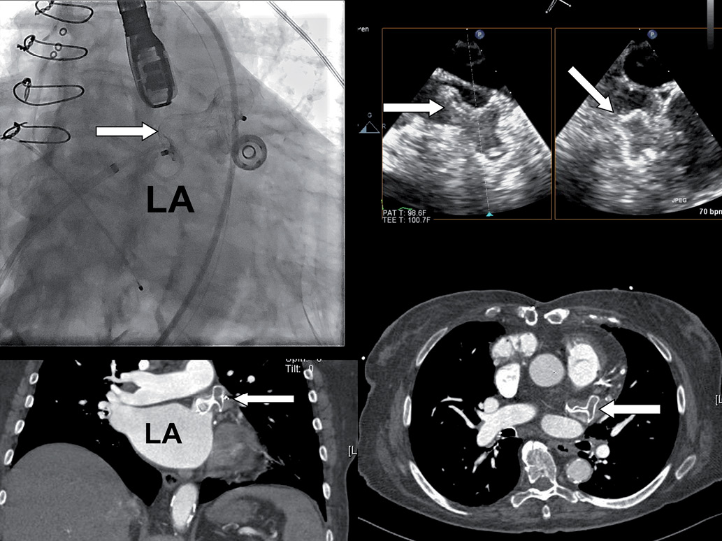
Figure 1. Left upper corner: fluoroscopic view of 20 mm septal occluder device after deployment in left atrial appendage. Right upper corner: biplane transoesophageal echocardiography images showing device positioned in left atrial appendage. Left lower corner: coronal view, and right lower quadrant: transverse view of post-procedure cardiac CT scan showing that the distal part of the device has assumed a cone shape conforming to the shape of the left atrial appendage and that there is no contrast enhancement of the appendage consistent with occlusion. Arrow points at the device. LA: left atrium
Conflict of interest statement
The authors have no conflicts of interest to declare.
Online data supplement
Moving image 1. Baseline left atrial appendage angiogram. Initial few beats demonstrate thrombus in left atrial appendage seen as contrast staining. Both the angiogram and live transoesophageal echocardiography were used to define the anatomy of left atrial appendage.
Moving image 2. Left atrial appendage thrombectomy. An Angiojet® catheter is seen in left atrial appendage. Mechanical thrombectomy is being performed.
Moving image 3. Final left atrial angiogram. The Amplatzer device is successfully deployed. There is residual thrombus in the appendage however contrast does not flow in all the way into the appendage (compared to baseline angiogram in Moving image 1) consistent with occlusion. A temporary pacemaker wire is also seen which was placed due to AV block during mechanical thrombectomy with Angiojet®.