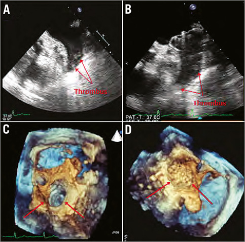
Occlusion of the left atrial appendage (LAA) using the WATCHMAN™ device (Boston Scientific, Natick, MA, USA) proved its safety and efficacy in the PROTECT-AF trial and has found its way into daily clinical routine since then. However, patients with known thrombus formation within the LAA have been excluded from this technique so far because of the high risk of embolisation during the procedure in these patients. We report the first two cases of successful LAA occlusion using the WATCHMAN™ device and cerebral protection systems in patients with known thrombus within the LAA. Both patients were successfully treated with a WATCHMAN™ device without procedural complications. As cerebral protection devices, a SpiderFX™ system (ev3 Endovascular, Inc., Plymouth, MN, USA) was used in the first case and a Claret Medical Montage™ (Claret Medical, Inc., Santa Rosa, CA, USA) for the second patient. We conclude that LAA occlusion with cerebral protection devices is feasible in selected patients with LAA thrombus at high risk for embolic complications.

Figure 1. Patient 1. A) Transoesophageal echocardiographic (TEE) view of the left atrial appendage (LAA) thrombus prior to implantation. B) WATCHMAN™ device after implantation with remaining thrombus in the distal LAA. C) 3D-TEE view of the LAA ostium with thrombus visible within the LAA. D) 3D-TEE after implantation of the WATCHMAN™ device.
Conflict of interest statement
The authors have no conflicts of interest to declare.
Online data supplement
Moving image 1. Angiographic view of the atypical approach for unfolding of the device used in the first patient. The delivery sheath is placed at the ostium of the left atrial appendage (LAA) and the device is unfolded by pushing the device forward into the LAA.
Moving image 2. Transoesophageal echocardiography after implantation of the WATCHMAN™ device in the first patient showing the device position and the thrombus further distal within the left atrial appendage.

Online Figure 1. Patient 2. A) Transoesophageal echocardiography (TEE) prior to implantation with thrombus and “smoke” within the left atrial appendage. B) Angiographic view of the Claret™ system placed in the left carotid artery and the brachiocephalic trunk. C) TEE view of the WATCHMAN™ device after implantation.
Supplementary data
To read the full content of this article, please download the PDF.
Moving image 1.
Moving image 2.

