A 57-year-old male patient with a history of a medically managed inferior myocardial infarction 10 years previously underwent invasive catheter angiography. Coronary angiography revealed a patent right coronary artery with TIMI 3 flow and multiple linear filling defects (Figure 1A). Intracoronary imaging was performed with a “Lunawave®” optical frequency domain imaging catheter (OFDI) (Terumo Corp., Tokyo, Japan). This revealed that the right coronary artery had recanalised with the presence of a multiply septated lumen (Figure 1B). The OFDI images underwent three-dimensional reconstruction, allowing visualisation of multiple large spiralling channels (Figure 1C). The development of neovascular channels in chronically occluded coronary arteries has been described in histological studies with diameters >250 µm considered large1. The spatial resolution of OFDI allows detailed in vivo assessment of coronary intima previously only possible with post mortem histology. We believe these are the first published three-dimensional OFDI images demonstrating the presence of multiple macrochannels, rather than microchannels, with diameters >1,000 µm separated by thin fibrous septa.
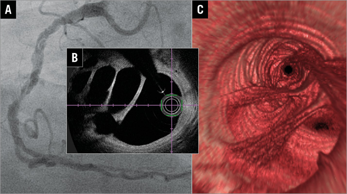
Figure 1. Coronary angiogram and OFDI imaging. A) Catheter angiography of right coronary artery (RCA) with multiple linear filling defects. B) OFDI of the mid RCA revealing multiple lumens. C) Three-dimensional reconstruction of OFDI showing recanalisation of the RCA with macrochannels and thin fibrous septa.
Conflict of interest statement
The authors have no conflicts of interest to declare.