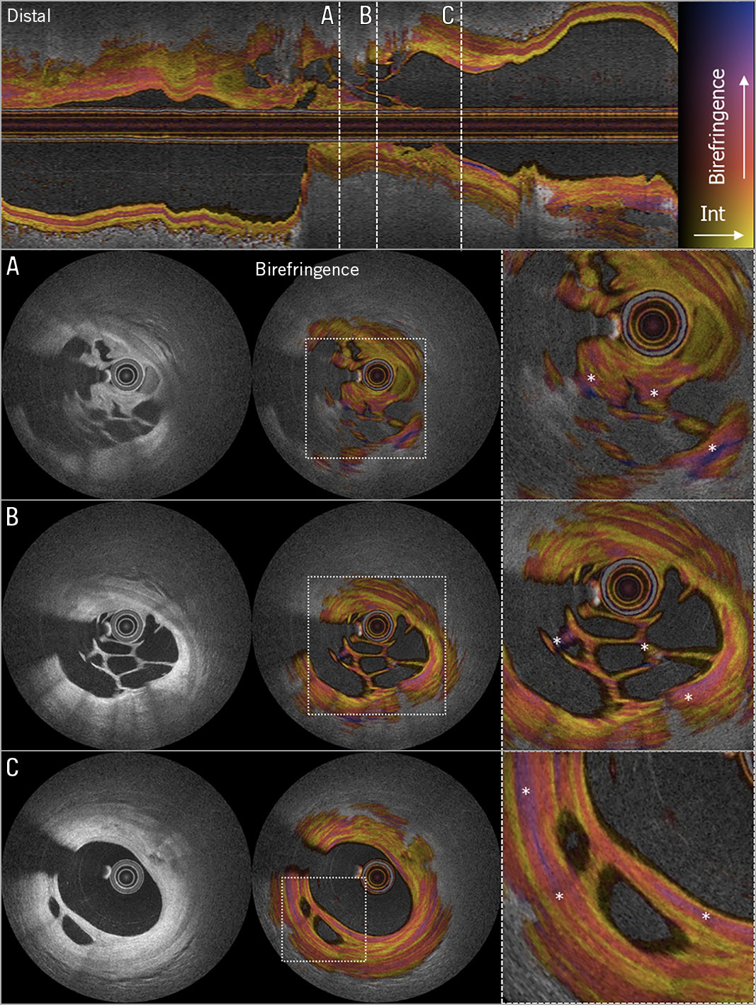
Figure 1. OFDI showing a honeycomb-like structure with multiple intraluminal microchannels confirming the presence of recanalised thrombus. Upper panel: longitudinal view of the right coronary artery. The dashed white lines denote the cross-sectional locations for A, B and C. A), B) & C) Three locations with clear recanalisation in organised thrombus. From left to right: conventional OFDI intensity, birefringence, magnified birefringence (* denotes zones of high birefringence surrounding microchannels).
A 69-year-old female patient was admitted to our institution for potential treatment of a non-ST-segment elevation myocardial infarction after eight days of intermittent pain. Angiography showed an unusual filling defect in the mid-right coronary artery with Thombolysis In Myocardial Infarction (TIMI) 3 flow and moderate stable disease in the left coronary system. Optical frequency domain imaging (OFDI) revealed a honeycomb-like structure with multiple intraluminal microchannels confirming the presence of recanalised thrombus (Figure 1). Fresh thrombus (<1 day) comprises platelet aggregates, erythrocytes, and fibrin. Lytic thrombus (1 to 5 days), containing areas of necrosis and granulocytes, transforms into organised thrombus (>5 days), characterised by the presence of smooth muscle cells (SMCs), homogeneous or hyalin fibrin, and depositions of connective tissue and capillary ingrowth1. The patient was included in the POLARIS-I registry, designed to assess the added value of measuring birefringence with polarisation-sensitive (PS)-OFDI in acute coronary syndrome (ACS) patients. Collagen and SMCs have been shown to display higher birefringence than other tissue components in the vessel wall2. In the present case (Figure 1, right panel), high birefringence signals can be appreciated in connective tissue between the microchannels, suggesting the presence of collagen and SMCs.
To date, the progression and consistency of thrombus can only be reliably assessed using histopathological examination, which has no place in an acute setting. PS-OFDI may be of value to study the age, stability and morphometric characteristics of coronary thrombus.
Funding
This work was supported by the National Institutes of Health (grants P41EB-015903 and R01HL-119065) and by Terumo Corporation.
Conflict of interest statement
B. Bouma was supported in part by the Professor Andries Querido visiting professorship of the Erasmus University Medical Center in Rotterdam. Massachusetts General Hospital and the Erasmus University Medical Center have patent licensing arrangements with Terumo Corporation. B. Bouma has the right to receive royalties as part of the licensing arrangements. J. Daemen reports having received institutional research support from Pie Medical, Acist Medical Inc., PulseCath, Medtronic, Boston Scientific, and Abbott Vascular, and speaker and consultancy fees from PulseCath, Medtronic, ReCor Medical, and Acist Medical Inc. K. Otsuka acknowledges partial support from the Japan Heart Foundation ̸ Bayer Yakuhin Research Grant Abroad, the Uehara Memorial Foundation Postdoctoral Fellowship, and the Japan Society for the Promotion of Science Overseas Research Fellowship. L. van Zandvoort has received institutional research support from Acist Medical Inc.
Supplementary data
To read the full content of this article, please download the PDF.