DOI:

Optical coherence tomography features of late-stage recanalised coronary thrombi

Periklis A. Davlouros*, MD, PhD; Vasileios Karantalis, MD; Eleni Mavronasiou, MD; Anastasia Damelou, MD; Dimitrios Alexopoulos, MD, PhD, FESC, FACC

Case description

Our figure displays optical coherence tomography (OCT), features of two cases with angiographically suspected recanalised thrombus.1

A 45-year-old smoking and hyperlipidaemic male was admitted to our hospital with an acute anterior myocardial infarction, successfully treated with primary angioplasty of the LAD. However, angiography also demonstrated an irregular intra-luminal contrast defect in the left circumflex (LCx) artery, (Panel A, black arrow). Three days later, OCT (M2 Cardiology Imaging System; LightLab Imaging, Inc., Westford, MA, USA), demonstrated (Panels B-F), signal rich, high-backscattering diaphragm-like protrusions (white arrows), dividing the lumen to small channels. In Panel F, red thrombus is depicted (asterisk).2 The lesion was stented successfully.

A 60-year-old male with a history of an old myocardial infarction was admitted to our hospital for coronary angiography due to stable angina with a positive exercise stress test. Panel G shows an 80% angiographic stenosis of the LCx with a filling defect, and luminal haziness, consistent with the angiographic features of a recanalised thrombus.1 OCT of the LCx (Panel H), shows signal rich, high-backscattering diaphragm-like protrusions, dividing the lumen to multiple small channels communicating with each other. Based on the angiographic appearance, we speculate that these diaphragm-like protrusions represent remnants of a late stage recanalised thrombus. The lesion was re-crossed without difficulty and stented successfully.

Complex angiographic filling defects may represent ruptured atheromatous plaques, coronary artery dissection, emboli, mural calcification, unapposed stent struts, or recanalised thrombus.3 In vivo clarification of the anatomy of such lesions has become possible with OCT.

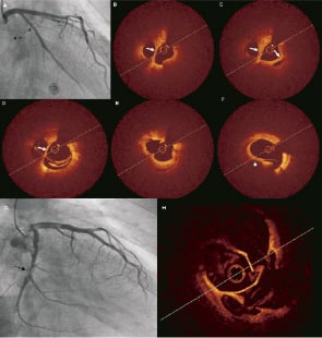
Figure 1. Angiographic and optical coherence tomography (OCT) findings in two patients (panels A-F, and G-H) with angiographically suspected recanalised thrombus.

Volume 6 Number 8
Mar 21, 2011
Volume 6 Number 8
View full issue


Key metrics

Suggested by Cory

Image – Interventional flashlight

10.4244/EIJ-D-20-00507 Aug 6, 2021
Optical coherence tomography and tailored treatment of in-stent restenosis
Jia H et al
free

CLINICAL RESEARCH

10.4244/EIJV11I3A59 Jul 20, 2015
Serial optical coherence tomography imaging of ACS-causing culprit plaques
Souteyrand G et al
free

Image – Interventional flashlight

10.4244/EIJ-D-20-00985 Aug 27, 2021
Distal embolisation as a cause of ST-segment elevation myocardial infarction: insights from optical coherence tomography
Satogami K et al
free

Image – Interventional flashlight

10.4244/EIJ-D-19-00431 Apr 20, 2021
Intracoronary polarimetry of a honeycomb-like structure
van Zandvoort L et al
free

Image – Interventional flashlight

10.4244/EIJ-D-22-00007 Aug 5, 2022
The fingerprints of plaque rupture healing as detected by serial optical coherence tomography imaging
Vergallo R et al
free

State-of-the-Art

10.4244/EIJ-D-21-00089 Jun 11, 2021
Intracoronary optical coherence tomography: state of the art and future directions
Ali ZA et al
free
Trending articles
224.5

State-of-the-Art Review

10.4244/EIJ-D-21-00426 Dec 3, 2021
Myocardial infarction with non-obstructive coronary artery disease
Lindahl B et al
free
172.9

Focus article

10.4244/EIJY19M08_01 Jan 17, 2020
EHRA/EAPCI expert consensus statement on catheter-based left atrial appendage occlusion – an update
Glikson M et al
free
96.2

State-of-the-Art

10.4244/EIJ-D-24-00066 Apr 21, 2025
Management of complications after valvular interventions
Bansal A et al
free
63

Image – Interventional flashlight

10.4244/EIJ-D-22-00074 Jun 3, 2022
Transcatheter aortic valve repair for aortic regurgitation with the Cusper device
Feld Y et al
free
58.2

Clinical research

10.4244/EIJ-D-23-00344 Sep 18, 2023
Clinical outcomes of TAVI with the Myval balloon-expandable valve for non-calcified aortic regurgitation
Sanchez-Luna JP et al
free
49.6

Original Research

10.4244/EIJ-D-25-00331 May 21, 2025
One-month dual antiplatelet therapy followed by prasugrel monotherapy at a reduced dose: the 4D-ACS randomised trial
Jang Y et al
open access
49.6

Original Research

10.4244/EIJ-D-25-00331 Jul 21, 2025
One-month dual antiplatelet therapy followed by prasugrel monotherapy at a reduced dose: the 4D-ACS randomised trial
Jang Y et al
open access
31.1

Expert Review

10.4244/EIJ-D-24-00535 May 5, 2025
Catheter-based techniques for pulmonary embolism treatment
Costa F et al
30.4

Clinical Research

10.4244/EIJ-D-21-00363 Jan 28, 2022
Membranous septum morphology and risk of conduction abnormalities after transcatheter aortic valve implantation
Jørgensen T et al
free
X

PCR
Impact factor: 9.5
2024 Journal Citation Reports®
Science Edition (Clarivate Analytics, 2025)
Online ISSN 1969-6213 - Print ISSN 1774-024X
© 2005-2025 Europa Group - All rights reserved