DOI: 10.4244/EIJV9I8A169

A unique feature of thin flat thrombus visualised by optical coherence tomography

Teruyoshi Kume, MD; Hiroyuki Okura*, MD; Kenzo Fukuhara, MD; Terumasa Koyama, MD; Shintaro Nezuo, MD; Yoji Neishi, MD; Akihiro Hayashida, MD; Takahiro Kawamoto, MD; Kiyoshi Yoshida, MD

Optical coherence tomography (OCT) has recently been developed as a high-resolution intravascular imaging modality, detecting small abnormal structures in the coronary lumen that could not be visualised by intravascular ultrasound (IVUS).

A 77-year-old man died with acute myelogenous leukemia. After he died, the coronary artery was imaged by OCT (ImageWire; LightLab Imaging, Westford, MA, USA) and IVUS (Atlantis SR Pro, 40 MHz; Boston Scientific, Natick, MA, USA). OCT clearly demonstrated a thin flat structure in the coronary lumen (Figure 1A, Moving image 1) but not in the IVUS image. This unique structural feature was demonstrated in corresponding histological images (Figure 1B, arrowhead). It revealed a white thrombus consisting mainly of platelets and white blood cells (Figure 1C and Figure 1D). This thin flat white thrombus was attached to the vessel wall without significant lumen stenosis.

High-resolution OCT was able to detect a unique feature of thrombus such as a thin flat structure in the coronary lumen. The clinical impact of this abnormal structure is still unclear. However, differentiation of intimal dissection might be necessary in the clinical setting.

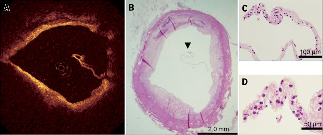
Figure 1. A thin flat thrombus visualised by optical coherence tomography. A thin flat structure in the coronary lumen was visualised by optical coherence tomography (A). Histological images revealed a white thrombus consisting mainly of platelets and white blood cells (B-D).

Conflict of interest statement

The authors have no conflicts of interest to declare.

Online data supplement

Moving image 1. OCT demonstrating a thin flat structure protruding into the coronary lumen during pullback.

Supplementary data

To read the full content of this article, please download the PDF.

Moving image 1. OCT demonstrating a thin flat structure protruding into the coronary lumen during pullback.

Volume 9 Number 8
Dec 27, 2013
Volume 9 Number 8
View full issue


Key metrics

Suggested by Cory

State-of-the-Art

10.4244/EIJ-D-21-00089 Jun 11, 2021
Intracoronary optical coherence tomography: state of the art and future directions
Ali ZA et al
free

Image – Interventional flashlight

10.4244/EIJ-D-19-00431 Apr 20, 2021
Intracoronary polarimetry of a honeycomb-like structure
van Zandvoort L et al
free

CLINICAL RESEARCH

10.4244/EIJV11I3A59 Jul 20, 2015
Serial optical coherence tomography imaging of ACS-causing culprit plaques
Souteyrand G et al
free

Image – Interventional flashlight

10.4244/EIJ-D-20-00985 Aug 27, 2021
Distal embolisation as a cause of ST-segment elevation myocardial infarction: insights from optical coherence tomography
Satogami K et al
free

Image – Interventional flashlight

10.4244/EIJ-D-20-00507 Aug 6, 2021
Optical coherence tomography and tailored treatment of in-stent restenosis
Jia H et al
free

Image – Interventional flashlight

10.4244/EIJ-D-21-00796 Jun 3, 2022
Patterns of intracoronary thrombus by high-definition intravascular ultrasound
Groenland F et al
free
Trending articles
309.93

State-of-the-Art Review

10.4244/EIJ-D-21-00695 Nov 19, 2021
Transcatheter treatment for tricuspid valve disease
Praz F et al
free
166.7

Expert review

10.4244/EIJ-D-21-00690 May 15, 2022
Crush techniques for percutaneous coronary intervention of bifurcation lesions
Moroni F et al
free
92.2

State-of-the-Art Review

10.4244/EIJ-D-20-01296 Aug 27, 2021
Management of cardiogenic shock
Thiele H et al
free
76

State-of-the-Art

10.4244/EIJ-D-23-00840 Sep 2, 2024
Aortic regurgitation: from mechanisms to management
Baumbach A et al
free
72.85

State-of-the-Art

10.4244/EIJ-D-24-00386 Feb 3, 2025
Mechanical circulatory support for complex, high-risk percutaneous coronary intervention
Ferro E et al
free
58.3

Clinical research

10.4244/EIJ-D-23-00344 Sep 18, 2023
Clinical outcomes of TAVI with the Myval balloon-expandable valve for non-calcified aortic regurgitation
Sanchez-Luna JP et al
free
56.65

Clinical research

10.4244/EIJ-D-20-01155 Oct 20, 2021
A deep learning algorithm for detecting acute myocardial infarction
Liu W et al
free
33.9

CLINICAL RESEARCH

10.4244/EIJ-D-17-00381 Oct 11, 2017
Stent malapposition and the risk of stent thrombosis: mechanistic insights from an in vitro model
Foin N et al
free
33.65

State-of-the-Art

10.4244/EIJ-D-23-00606 Jan 1, 2024
Targeting inflammation in atherosclerosis: overview, strategy and directions
Waksman R et al
free
X

The Official Journal of EuroPCR and the European Association of Percutaneous Cardiovascular Interventions (EAPCI)

EuroPCR EAPCI
PCR ESC
Impact factor: 7.6
2023 Journal Citation Reports®
Science Edition (Clarivate Analytics, 2024)
Online ISSN 1969-6213 - Print ISSN 1774-024X
© 2005-2025 Europa Group - All rights reserved