IMAGE IN CARDIOLOGY

DOI: 10.4244/EIJV12I1A20

Intracardiac thrombus trapped in a patent foramen ovale

Oezguer A. Onur1,2*, MD; Ferdinand Kuhn-Régnier3, MD; Christoph Kabbasch4, MD; Gereon R. Fink1,2, MD; Jochen Müller-Ehmsen5, MD

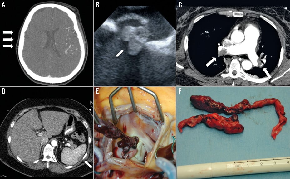
A 57-year-old woman was admitted to the University Hospital of Cologne after her husband had found her unconscious. Upon admission, gaze deviation to the right was observed. A cerebral CT scan revealed right hemispheric hypoperfusion (Panel A) and thrombolysis was performed. Subsequently, transoesophageal echocardiography showed a thrombus, which was trapped in a patent foramen ovale (Panel B, Moving image 1). A CT scan of the thorax and the abdomen revealed multiple pulmonary embolisms and an infarction of the spleen (Panel C, Panel D). A thrombectomy of the thrombus, with a length of 12 cm, and a closure of the patent foramen ovale were performed (Panel E, Panel F). Follow-up imaging revealed only a small cerebral infarct. Neurological examination was inconspicuous.

A consent form was obtained from the patient even though she cannot be personally identified from the images contained in this article.

Conflict of interest statement

The authors have no conflicts of interest to declare.

Supplementary data

Moving image 1. Thrombus trapped in a patent foramen ovale. Transoesophageal echocardiography showing a thrombus (arrowheads) in the right atrium, trapped in a patent foramen ovale (arrow), and extending into the left atrium. LA: left atrium; RA: right atrium
Supplementary data

To read the full content of this article, please download the PDF.

Thrombus trapped in a patent foramen ovale. Transoesophageal echocardiography showing a thrombus (arrowheads) in the right atrium, trapped in a patent foramen ovale (arrow), and extending into the left atrium. LA: left atrium; RA: right atrium

Volume 12 Number 1
May 16, 2016
Volume 12 Number 1
View full issue


Key metrics

Suggested by Cory

10.4244/EIJV12SXA2 May 16, 2016
Patent foramen ovale: indications for closure and techniques
Taramasso M et al
free

Viewpoint

10.4244/EIJ-D-21-00500 Oct 20, 2021
My stroke story
Noc M
free

IMAGE IN CARDIOLOGY

10.4244/EIJY14M10_04 Oct 20, 2015
An unusual complication of hip surgery: paradoxical coronary embolism of foreign material as a cause of acute MI
Mallouppas M et al
free

Editorial

10.4244/EIJ-E-24-00040 Aug 19, 2024
The patent foramen ovale does not spontaneously close in patients aged 60
Hildick-Smith D and Arunothayaraj S
free

10.4244/EIJV14I13A244 Jan 18, 2019
Patent foramen ovale: the known unknowns
Thaler D and Gans S
free

CLINICAL RESEARCH

10.4244/EIJV10I12A257 Apr 20, 2015
Very long-term follow-up after percutaneous closure of patent foramen ovale
Eeckhout E et al
free

IMAGE IN CARDIOLOGY

10.4244/EIJV10I7A146 Nov 20, 2014
Percutaneous reclosure of a patent foramen ovale after bioabsorbable device implantation
Snijder R et al
free
Trending articles
151.43

State-of-the-Art

10.4244/EIJ-D-22-00776 Apr 3, 2023
Computed tomographic angiography in coronary artery disease
Serruys PW et al
free
55.9

Clinical research

10.4244/EIJ-D-22-00621 Feb 20, 2023
Long-term changes in coronary physiology after aortic valve replacement
Sabbah M et al
free
54.9

Expert review

10.4244/EIJ-D-21-01010 Jun 24, 2022
Device-related thrombus following left atrial appendage occlusion
Simard T et al
free
43.75

Clinical Research

10.4244/EIJ-D-21-01091 Aug 5, 2022
Lifetime management of patients with symptomatic severe aortic stenosis: a computed tomography simulation study
Medranda G et al
free
39.95

Clinical research

10.4244/EIJ-D-22-00558 Feb 6, 2023
Permanent pacemaker implantation and left bundle branch block with self-expanding valves – a SCOPE 2 subanalysis
Pellegrini C et al
free
X

The Official Journal of EuroPCR and the European Association of Percutaneous Cardiovascular Interventions (EAPCI)

EuroPCR EAPCI
PCR ESC
Impact factor: 7.6
2023 Journal Citation Reports®
Science Edition (Clarivate Analytics, 2024)
Online ISSN 1969-6213 - Print ISSN 1774-024X
© 2005-2024 Europa Group - All rights reserved