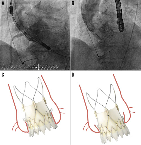

An 82-year-old man with multiple comorbidities had severe symptomatic aortic regurgitation due to degeneration of the leaflets and prolapse of the right coronary cusp (Moving image 1). Computed tomography revealed an annulus of 22×28 mm (mean 25 mm) without relevant calcification. We hypothesised that an ACURATE neo™, L (Symetis SA, Ecublens, Switzerland) transcatheter heart valve (THV) might provide enough oversizing to anchor the valve securely within the annulus, despite the absence of calcification. Bail-out open heart surgery was discussed with the patient in case of procedural failure.
To improve visualisation during transcatheter aortic valve implantation, positioning and deployment of the THV was performed under rapid pacing (Panel A, Panel B, Moving image 2- Moving image 4). Within the first minutes after implantation, the valve migrated a few millimetres towards the left ventricle, until it had reached a stable position with the upper crown sitting just above the native valve leaflets (Panel C, the initial position after release, Panel D, the final position). Since the amount of migration was very limited, there was still sufficient sealing with the native annulus (Moving image 5, Moving image 6). In this case, the upper crown of the ACURATE neo (L), the outer diameter of which is about 32 mm, was crucial to anchor the valve and prevent it from further migration into the left ventricle. In conclusion, the Symetis ACURATE neo transfemoral THV may be well suited for the treatment of uncalcified, pure aortic regurgitation, as long as there is the option for sufficient oversizing.
Conflict of interest statement
S. Toggweiler is a proctor for Symetis. The other authors have no conflicts of interest to declare.
Supplementary data
Moving image 1. Baseline echocardiography of the aortic regurgitation.
Moving image 2. Initial positioning of the ACURATE neo under semi-rapid pacing.
Moving image 3. Positioning after deployment of the upper crown under rapid pacing.
Moving image 4. Full deployment of the valve prosthesis under rapid pacing.
Moving image 5. Final angiogram.
Moving image 6. Transthoracic echocardiography at one-month follow-up.
Supplementary data
To read the full content of this article, please download the PDF.
Baseline echocardiography of the aortic regurgitation.
Initial positioning of the ACURATE neo under semi-rapid pacing.
Positioning after deployment of the upper crown under rapid pacing.
Full deployment of the valve prosthesis under rapid pacing.
Final angiogram.
Transthoracic echocardiography at one-month follow-up.

