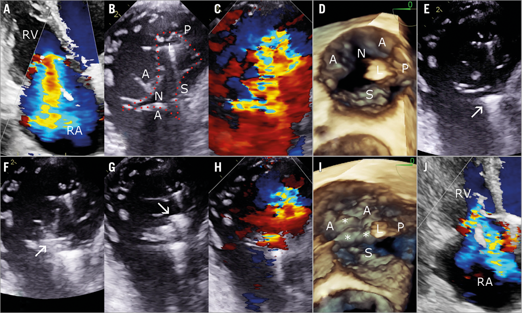
An 80-year-old woman presented with refractory right heart failure, recurrent hospitalisations and massive secondary tricuspid regurgitation (TR). She had persistent NYHA Class IV dyspnoea, severe fatigue and peripheral oedema. Past history included atrial fibrillation, dual chamber pacemaker five years prior, coronary disease, transient ischaemic attack, hypertension and chronic kidney disease. Echocardiography demonstrated massive secondary TR predominantly due to annular dilatation, leaflet tethering and a large notch in the anterior leaflet such that the valve was functionally quadricuspid (Panel A-Panel D, Moving image 1-Moving image 3). The pacemaker lead was located in the posteroseptal commissure and did not appear to be the main cause of TR. After Heart Team review, the patient was accepted for transcatheter repair with the MitraClip® (Abbott Vascular, Santa Clara, CA, USA).
The first clip was deployed between the anterior and septal leaflets near the commissure (Panel E, Moving image 4, Moving image 5). The anterior leaflet notch was then treated, placing the second clip perpendicular to the first clip within the notch, effectively sealing it during systole (Panel F, Moving image 6, Moving image 7). This also brought the anterior and septal leaflets closer together, allowing implantation of a third clip near the centre of the valve, reducing TR from massive to moderate, with a mean gradient of 2 mmHg (Panel G-Panel J, Moving image 8-Moving image 11). The patient’s condition improved significantly, allowing hospital discharge. At one-month follow-up, the patient reported improved symptoms with reduced diuretic requirements. Echocardiography demonstrated sustained TR reduction.
The tricuspid valve is a dynamic structure that changes with loading conditions. Although true congenital tricuspid clefts are rare1, notches of the anterior and septal leaflets are common on direct inspection of pathologic specimens2,3. Tricuspid notches are probably not recognised on routine echocardiographic imaging protocols, but are increasingly seen in patients referred for tricuspid intervention. With progressive RV remodelling and annular dilatation in secondary TR, these notches may become stretched open, further contributing to TR progression. In this first report, we describe successful treatment of a large anterior leaflet notch with the MitraClip, leading to improved leaflet coaptation and sustained TR reduction with significant clinical improvement. This approach parallels the surgical technique of suture-based closure of large notches causing TR3. As interventional imaging of the tricuspid valve evolves, increased recognition of tricuspid notches will be important for successful treatment strategy and clinical outcomes.
Conflict of interest statement
The authors have no conflicts of interest to declare.
Supplementary data
Moving image 1. Preprocedural TTE RV-focused apical view demonstrating massive TR.
Moving image 2. Preprocedural TEE transgastric view of the tricuspid valve with simultaneous colour Doppler demonstrating massive TR and a large anterior leaflet notch.
Moving image 3. Preprocedural 3D TEE demonstrating anterior leaflet notch and pacemaker lead position.
Moving image 4. Periprocedural TEE transgastric view of grasping anterior and septal leaflets near the commissure.
Moving image 5. Periprocedural TEE transgastric view after implantation of one clip between anterior and septal leaflets, with persistence of the anterior leaflet notch.
Moving image 6. Periprocedural TEE transgastric view of grasping within the anterior leaflet notch, perpendicular to the first clip.
Moving image 7. Periprocedural TEE transgastric view after implantation of a second clip across the anterior leaflet notch, showing a subsequent change in valve leaflet motion and closure of the notch during systole.
Moving image 8. Periprocedural TEE transgastric view after implantation of a third clip between anterior and septal leaflets near the middle of the valve.
Moving image 9. Post-procedural TEE transgastric view with colour Doppler demonstrating residual moderate TR.
Moving image 10. Post-procedural 3D TEE after implantation of three clips.
Moving image 11. Post-procedural TTE RV-focused apical view demonstrating residual moderate TR.
Supplementary data
To read the full content of this article, please download the PDF.
Post-procedural TEE transgastric view with colour Doppler demonstrating residual moderate TR.
Preprocedural TTE RV-focused apical view demonstrating massive TR.
Post-procedural 3D TEE after implantation of three clips.
Post-procedural TTE RV-focused apical view demonstrating residual moderate TR.
Preprocedural TEE transgastric view of the tricuspid valve with simultaneous colour Doppler demonstrating massive TR and a large anterior leaflet notch.
Preprocedural 3D TEE demonstrating anterior leaflet notch and pacemaker lead position.
Periprocedural TEE transgastric view of grasping anterior and septal leaflets near the commissure.
Periprocedural TEE transgastric view after implantation of one clip between anterior and septal leaflets, with persistence of the anterior leaflet notch.
Periprocedural TEE transgastric view of grasping within the anterior leaflet notch, perpendicular to the first clip.
Periprocedural TEE transgastric view after implantation of a second clip across the anterior leaflet notch, showing a subsequent change in valve leaflet motion and closure of the notch during systole.
Periprocedural TEE transgastric view after implantation of a third clip between anterior and septal leaflets near the middle of the valve.