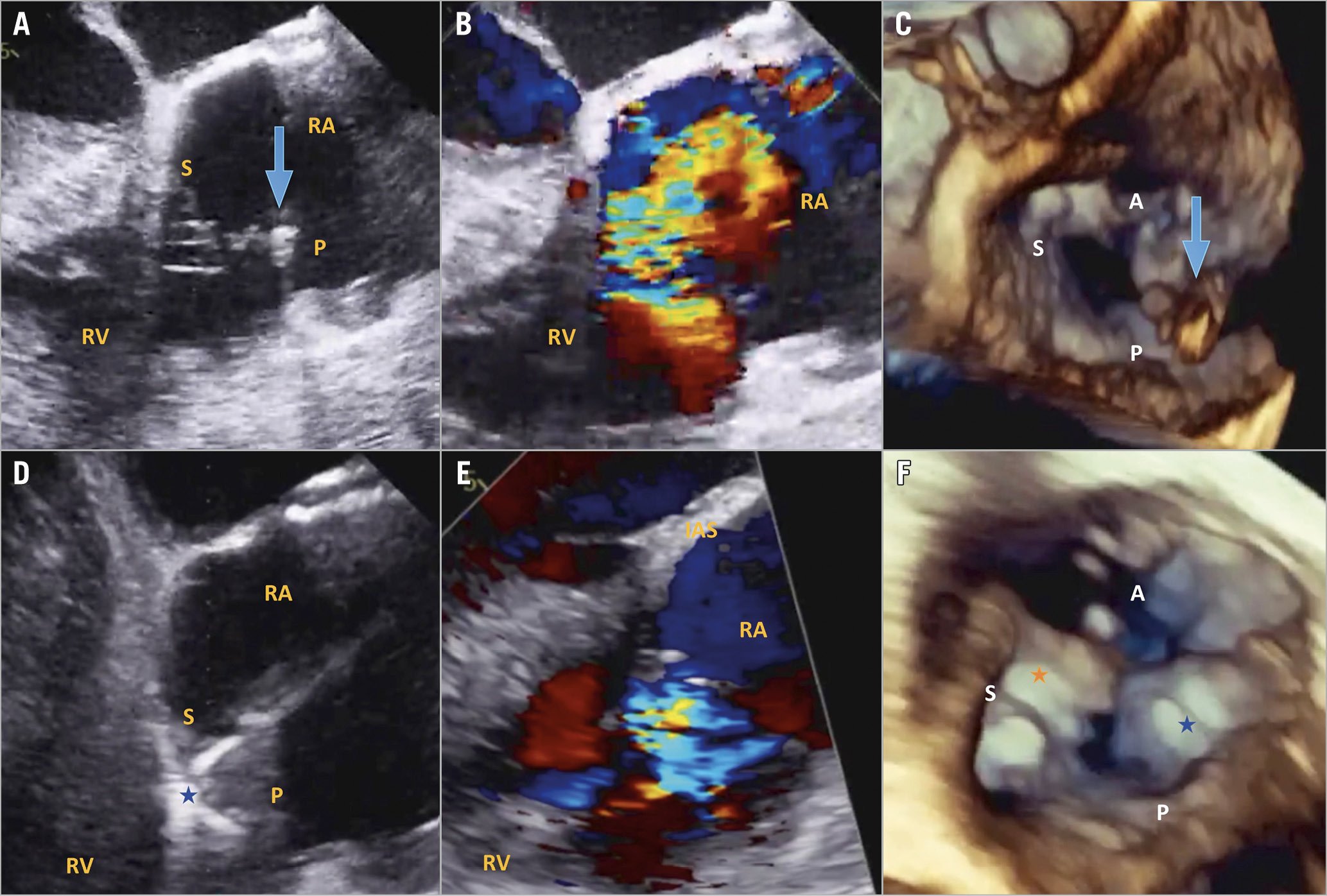
Figure 1. TTVr with the modified TriClip/MitraClip G4 system. A) Baseline transoesophageal echo (TEE) 150-degree view demonstrating large posterior leaflet flail. B) Baseline TEE 150-degree view with colour Doppler demonstrating torrential tricuspid regurgitation (TR). C) 3D TEE view demonstrating posterior leaflet flail. D) TEE 150-degree view of leaflet grasping with the MitraClip G4 XTW. E) Post-procedural TEE 150-degree view demonstrating moderate TR. F) 3D TEE view demonstrating tissue bridge and elimination of posterior flail. A: anterior leaflet; P: posterior leaflet; RA: right atrium; RV: right ventricle; S: septal leaflet. Blue arrow indicates flail posterior leaflet, orange star indicates TriClip XT between anterior and septal leaflets, blue star indicates MitraClip G4 XTW between posterior and septal leaflets.
A 57-year-old woman presented with progressive New York Heart Association (NYHA) Class III dyspnoea, oedema and fatigue. Past history included hypertrophic cardiomyopathy with recent implantable cardioverter-defibrillator (ICD) lead extraction due to high impedance, followed by subcutaneous ICD implantation. Echocardiography subsequently demonstrated torrential tricuspid regurgitation (TR) with posterior leaflet flail and a 15 mm flail gap (Figure 1A-Figure 1C, Moving image 1-Moving image 3). The right ventricle (RV) was dilated with normal function; left ventricular (LV) function was preserved with normal pulmonary pressures. After Heart Team review, the patient underwent transcatheter tricuspid valve repair (TTVr) with the modified TriClip™/MitraClip™ G4 system (Abbott Vascular, Santa Clara, CA, USA).
Initial attempts to treat the posterior leaflet flail using a TriClip XT clip were unsuccessful due to the large flail gap. Instead, a TriClip XT clip was placed centrally between the anterior and septal leaflets. Subsequently, a MitraClip G4 XTW clip was inserted into the TriClip guide without miskeying and steered down to the tricuspid valve using the P and L knobs. Using the wider clip, the posterior and septal leaflets were successfully grasped simultaneously, eliminating the flail segment and reducing TR from torrential to moderate (Figure 1D-Figure 1F, Moving image 4-Moving image 6). The patient had an excellent clinical response with improved dyspnoea and was discharged the following day. At one-month follow-up, the patient was in NYHA Class II and echocardiography demonstrated normal RV function with moderate TR, mean gradient 2 mmHg (Moving image 7).
TTVr has recently emerged as a safe and efficacious intervention for patients with severe TR and heart failure1. Worldwide, the majority of TTVr cases have utilised MitraClip NT or XT off-label using a modified steering technique2. The recently introduced TriClip system utilises a dedicated guide catheter with improved steering mechanism to facilitate a coaxial approach to the tricuspid valve. The MitraClip G4 XTW is 50% wider than the XT clip and allows independent leaflet grasping. Here, we implanted a MitraClip G4 XTW off-label using the TriClip guide and a modified steering technique, allowing successful treatment of a patient with challenging tricuspid anatomy. Further device iteration combined with procedural innovation may expand the number of patients eligible for TTVr.
Conflict of interest statement
N. Fam has received speaker honoraria from Abbott Vascular and is a consultant for Edwards Lifesciences. The other authors have no conflicts of interest to declare.
Supplementary data
To read the full content of this article, please download the PDF.
Moving image 1. Baseline echo demonstrating large posterior leaflet flail.
Moving image 2. Baseline echo demonstrating torrential TR.
Moving image 3. 3D echo demonstrating posterior leaflet flail.
Moving image 5. Post-procedural echo demonstrating moderate TR.
Moving image 6. 3D echo demonstrating tissue bridge and elimination of posterior flail.
Moving image 7. Echo at one-month follow-up demonstrating moderate TR.