IMAGE IN CARDIOLOGY

DOI: 10.4244/EIJV11I2A38

Cholesterol crystals identified using optical coherence tomography and virtual histology intravascular ultrasound

Adam J. Brown1,2*, MD; Daniel R. Obaid1, MD, PhD; Nick E. J. West2, MD; Martin Goddard3, MD; Martin R. Bennett1, MD, PhD

The deposition and accumulation of lipid within the arterial wall leads to the development and progression of atherosclerotic plaques. Cholesterol crystals (CCs) are formed from these lipid pools and both augment the inflammatory response and promote plaque rupture, possibly through inducing mechanical instability. Accordingly, identification of these structures is important for plaque risk stratification.

Optical coherence tomography (OCT) and virtual histology intravascular ultrasound (VH-IVUS) are invasive imaging modalities that permit plaque characterisation. Validation studies using these techniques have demonstrated their reliability to identify plaque constituents, including lipid and calcification. CCs are thought to be identifiable on OCT as linear, high-backscattering structures within fibroatheroma. However, OCT validation studies on CCs are lacking and their appearance on VH-IVUS unknown.

Here we present co-registered OCT (C7 Dragonfly™; St. Jude Medical, St. Paul, MN, USA) and VH-IVUS (Eagle Eye® Gold; Volcano Corporation, Rancho Cordova, CA, USA) images obtained from a human coronary artery imaged ex vivo on a proprietary pressure-perfused imaging rig (Figure 1). Consistent with previous experience, CCs (Figure 1A, Figure 1B) were clearly identified by OCT as bright, high-backscattering structures located within a lipid-rich plaque (Figure 1C). In contrast, on co-registered IVUS imaging, CCs resulted in a region of echogenic reflectivity (Figure 1D), which led to them being characterised as dense calcium by VH-IVUS spectral analysis (Figure 1E).

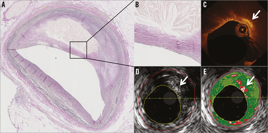
Figure 1. Co-registered images demonstrating cholesterol crystals on both optical coherence tomography and virtual histology intravascular ultrasound. An advanced atherosclerotic coronary lesion visualised by histology (A and B) with evidence of cholesterol crystals (arrows), identified by optical coherence tomography (C), greyscale intravascular ultrasound (D) and virtual histology intravascular ultrasound (E).

These images suggest that OCT offers improved diagnostic plaque characterisation compared with VH-IVUS for identification of cholesterol crystals in plaques.

Conflict of interest statement

The authors have no conflicts of interest to declare.

Volume 11 Number 2
Jun 19, 2015
Volume 11 Number 2
View full issue


Key metrics

Suggested by Cory

10.4244/EIJV16I5A66 Aug 28, 2020
Vulnerable plaque imaging – a clinical reality?
Johnson T and Joshi N
free

Preclinical research

10.4244/EIJ-D-20-00202 Aug 28, 2020
Detection of cholesterol crystals by optical coherence tomography
Jinnouchi H et al
free

State-of-the-Art

10.4244/EIJ-D-24-00387 Jul 21, 2025
Advances in coronary imaging of atherosclerotic plaques
Garcia-Garcia H et al
free

CLINICAL RESEARCH

10.4244/EIJ-D-16-00317 Jun 20, 2017
Limitations of OCT in identifying and quantifying lipid components: an in vivo comparison study with IVUS-NIRS
Di Vito L et al
free

Image – Interventional flashlight

10.4244/EIJ-D-19-00775 Apr 2, 2021
Correlation of near-infrared spectroscopy, optical coherence tomography and post-mortem histology for lipid detection
Zanchin C et al
free

State-of-the-Art

10.4244/EIJ-D-21-00089 Jun 11, 2021
Intracoronary optical coherence tomography: state of the art and future directions
Ali ZA et al
free

10.4244/EIJV8I1A3 May 15, 2012
When the doctor needs an engineer to be the matchmaker
Patel P and Chen Z
free
Trending articles
87.2

State-of-the-Art

10.4244/EIJ-D-25-00266 Jan 19, 2026
Lesion stratification with intracoronary imaging
McGarvey M et al
free
47.45

NEW INNOVATION

10.4244/EIJ-D-15-00467 Feb 20, 2018
Design and principle of operation of the HeartMate PHP (percutaneous heart pump)
Van Mieghem NM et al
free
34.8

Original Research

10.4244/EIJ-D-25-01006 Mar 16, 2026
Clinical outcomes and haemodynamic response after blinded stress assessment of moderate aortic stenosis
Eerdekens R et al
22.2

Viewpoint

10.4244/EIJ-D-25-01066 May 4, 2026
Intracoronary imaging guidance for de novo coronary lesion treatment with drug-coated balloons
Amabile N et al
free
20.75

Flashlight

10.4244/EIJ-D-25-01014 Apr 6, 2026
Stent retriever-assisted coronary thrombectomy with continuous aspiration
Liabot Q et al
open access
19.5

Original Research

10.4244/EIJ-D-26-00032 May 15, 2026
Glucocorticoids to reduce permanent pacemaker implantation after TAVI: the GLUCO-TAVI randomised trial
Fuertes-Kenneally L et al
17.8

Expert Review

10.4244/EIJ-D-25-01316 Apr 20, 2026
Electrosurgical laceration and stabilisation of tricuspid edge-to-edge repair: the ELASTA-T technique
Alvarez-Covarrubias H et al
free
X

PCR
Impact factor: 9.5
2024 Journal Citation Reports®
Science Edition (Clarivate Analytics, 2025)
Online ISSN 1969-6213 - Print ISSN 1774-024X
© 2005-2026 Europa Group - All rights reserved