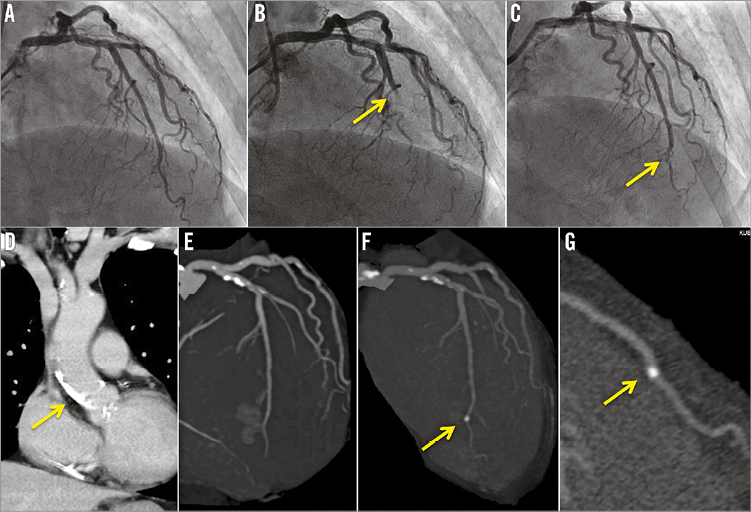

A 65-year-old female was admitted to our hospital for percutaneous coronary intervention (PCI) for chronic total occlusion (CTO) of the right coronary artery (RCA). Coronary angiography showed a CTO lesion in the mid RCA with good collateral circulation supplied from the left coronary artery (LCA) without significant stenosis or calcification (Panel A, Moving image 1). However, after simultaneous angiography, the patient had sudden chest pain with ST-elevation in leads V1-4. LCA angiography showed total occlusion in the mid left anterior descending coronary artery (LAD) (Panel B, Moving image 2). Emergent PCI of the LAD was then undertaken. While thrombus aspiration was tried repeatedly, no thrombus was aspirated, but the occluded site moved from mid to distal LAD (Moving image 3). After dilation with a 1.25 mm balloon, the occluded lesion improved to mild stenosis (Panel C, Moving image 4) and the patient’s symptoms disappeared. Computed tomography (CT) before PCI revealed severe calcification at the ascending aorta and no calcified finding at the distal LAD (Panel D, Panel E). Moreover, coronary CT angiography after PCI revealed a new calcified finding in accordance with the mild stenotic lesion of the distal LAD (Panel F, Panel G). We speculate that the calcified plaque of the ascending aorta embolised at the mid LAD during the catheter manipulation in the LCA.
Conflict of interest statement
The authors have no conflicts of interest to declare.
Supplementary data
Moving image 1. The initial LCA angiography showed no significant stenosis.
Moving image 2. The mid LAD was totally occluded after simultaneous angiography.
Moving image 3. After aspiration was tried repeatedly, the occluded site moved from mid to distal LAD.
Moving image 4. Finally, the mild stenosis remained at the distal LAD.
Supplementary data
To read the full content of this article, please download the PDF.
Moving image 1. The initial LCA angiography showed no significant stenosis.
Moving image 2. The mid LAD was totally occluded after simultaneous angiography.
Moving image 3. After aspiration was tried repeatedly, the occluded site moved from mid to distal LAD.
Moving image 4. Finally, the mild stenosis remained at the distal LAD.

