We report two cases of aortic valve stent thrombosis after uneventful transcatheter transapical aortic valve implantation of the 29 mm Edwards SAPIEN XT (Edwards Lifesciences, Irvine, CA, USA) in high-risk patients. Subcutaneous low molecular weight heparin 5,000 units daily for five days, aspirin 75 mg daily lifelong, and clopidogrel 75 mg daily for 12 months were given. After four weeks, antiplatelet therapy was reduced to monotherapy due to bleeding problems. Shortly after, the patients developed refractory heart failure and died on day 106 and on day 137, respectively. Transoesophageal echocardiography (Online Figure 1 and Moving image 1 and 2) and multislice computed tomography (Figure 1A and Figure 1B) showed signs of stent valve thrombosis.
The autopsy and histology showed that the valve leaflets were thickened, stiff and with fibrosis on both sides of the leaflets, which was more than three months old. Two layers of thrombosis were seen on top of the fibrosis (Figure 1C and Figure 1D). The most profound layer of thrombus was more than one month old, and the fresh thrombosis was very recent with the specific lines of Zahn. There were no signs of macro defects or endocarditis on the leaflets.
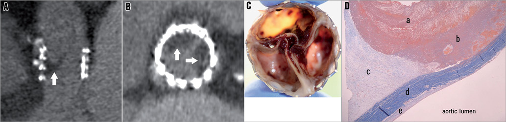
Figure 1. A) Multislice computed tomography image in case 1 showing the aortic stent prosthesis in the long-axis view in systole with thrombus (low attenuation mass, arrow) and fixation of the “right” leaflet. B) Multislice computed tomography image in case 1 showing the aortic stent prosthesis in short-axis view in systole with thrombus visualised as low attenuation masses (arrows) in relation to the “right” and “left” leaflet, respectively. C) Macroscopic image of the aortic valve stent. D) Masson trichrome stained section of a thrombosed leaflet: a) fresh thrombosis, b) recent thrombosis, c) fibrosis, d) native leaflet, and e) fibrosis.
Conflict of interest statement
The authors have no conflicts of interest to declare.
Online data supplement
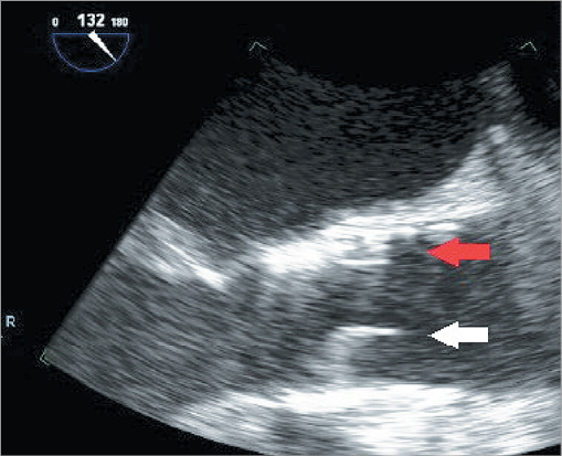
Online Figure 1. Transoesophageal echocardiography in case 1 showing the aortic stent prosthesis in the long-axis view with an immobile “right” valve leaflet (white arrow) whereas the “non-coronary” leaflet (red arrow) is well opened in systole.
Moving image 1. Transoesophageal echocardiogram in case 2 showing the aortic stent prosthesis in the two-dimensional longitudinal axis.
Moving image 2. The three-dimensional short-axis view with a thickened and immobile “right” valve leaflet.
Supplementary data
To read the full content of this article, please download the PDF.
Moving image 1. Transoesophageal echocardiogram in case 2 showing the aortic stent prosthesis in the two-dimensional longitudinal axis.
Moving image 2. The three-dimensional short-axis view with a thickened and immobile "right" valve leaflet.