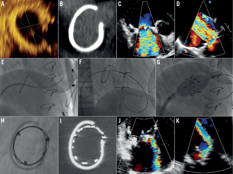
A 44-year-old man had twice undergone tricuspid valve replacement for endocarditis, using a mitral homograft and a 30 mm tricuspid Carpentier-Edwards Classic annuloplasty ring (Edwards Lifesciences, Irvine, CA, USA) (Figure 1A, Figure 1B) during the last operation in 2001. Endocarditis recurrences resulted in massive tricuspid regurgitation (TR), (Figure 1C, Figure 1D; Moving image 1, Moving image 2) with progressive right-sided heart failure and dyspnoea. Due to the high risk of further surgery, transfemoral tricuspid valve-in-ring implantation of a 26 mm Edwards SAPIEN XT® (Edwards Lifesciences) valve was performed (Figure 1E-Figure 1I; Moving image 3). There was a moderate residual paravalvular leak (Figure 1J, Figure 1K; Moving image 4, Moving image 5). Three months later, the TR was mild, and the patient was free of symptoms.

Figure 1. Tricuspid ring by 3D transoesophageal echocardiography (TOE) (A) and CT scan (B). Transthoracic echocardiography (TTE) (C) and TOE (D) showing massive TR. (E) Crossing of the tricuspid valve with a balloon floatation catheter on fluoroscopy. (F) Guidewire placed in the right ventricle. (G) Implantation of the SAPIEN XT valve. Fluoroscopy (H) and CT (I) of the implanted valve. TTE (J) and TOE (K) showing moderate residual paravalvular regurgitation.
Conflict of interest statement
D. Himbert is a proctor for Edwards Lifesciences. A. Vahanian receives speaker’s fees/honoraria from Edwards Lifesciences. The other authors have no conflicts of interest to declare.
Online data supplement
Moving image 1. Transthoracic echocardiography (TTE) showing a massive TR.
Moving image 2. TOE showing a massive TR.
Moving image 3. Implantation of the SAPIEN XT prosthesis in the tricuspid ring.
Moving image 4. TTE showing a moderate residual paravalvular TR.
Moving image 5. TOE showing a moderate residual paravalvular TR.
Supplementary data
To read the full content of this article, please download the PDF.
Moving image 1. Transthoracic echocardiography (TTE) showing a massive TR.
Moving image 2. TOE showing a massive TR.
Moving image 3. Implantation of the SAPIEN XT prosthesis in the tricuspid ring.
Moving image 4. TTE showing a moderate residual paravalvular TR.
Moving image 5. Transthoracic echocardiography (TTE) showing a massive TR.Moving image 2. TOE showing a massive TR.Moving image 3. Implantation of the SAPIEN XT prosthesis in the tricuspid ring. TTE showing a moderate residual paravalvular TR.Moving image 5. TOE showing a moderate residual paravalvular TR.

