Introduction
In this chapter of Tools & Techniques we discuss key points for successful stent delivery in difficult situations. While many techniques of stent delivery have been developed for these situations, still, difficulties in stent delivery may be encountered due to vessel tortuosity and/or calcification. The following is an introduction and summary of the considerations involved in stent delivery including specific techniques such as distal balloon deflection and distal balloon deflation techniques. The complete, unabridged e-version with dynamic images can be viewed at www.eurointervention.org.
Background
Stent delivery failure has been reported to occur in 3.7% of all cases and 92% of these failures are due to vessel tortuosity and/or calcification1. Another report shows that nearly 5% of lesions could not be successfully treated with the drug-eluting stents (DES)2.
Difficulties
When a stent fails to proceed forward because of severe tortuosity and/or calcification of the proximal section, it is imperative to assess the stability of the guide position, the stiffness of the wire, the calcification of the vessel and the flexibility of the stent. In clinical practice, multiple techniques such as proximal vessel preparation with balloon angioplasty or debulking techniques, vessel straightening with extra support and buddy wires, the use of alternative guiding catheters, or the increase of guiding catheter support by deep intubation using anchoring techniques or by the use of an inner catheter may be helpful to overcome such situations.
For each of these situations there are particular issues regarding equipment selection and techniques required for stent delivery which are discussed in the online version along with suggested tips and tricks to solve specific problems.
Techniques
Some of these techniques are rather easy, such as “deep breathing”, which will make the heart move more vertically, elongating the coronary vessel. This vessel straightening and elongation is often enough to enable the operator to move the stent forward into the vessel. Sometimes the guiding catheter support can be simply increased by a coaxial alignment of the catheter with the vessel. This can be achieved, e.g., by clockwise rotation for the right coronary artery (RCA) and the left circumflex coronary artery (LCX), or by counter-clockwise rotation for the left anterior descending coronary artery (LAD).
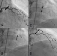
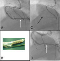
Other techniques require skilled manipulations of the guidewire, such as the coronary wire bias method, the use of support and buddy wires, or the jailing of coronary wires by stents and balloons. Sometimes it is necessary to modify the proximal section of the vessel or the lesion by predilatation, or with debulking techniques. Sometimes elaborate stent delivery techniques, such as the partial inflation of a deployed stent in order to cross a tortuous segment, are employed where the use of 5 or 4 Fr inner catheters (Figure 1), or of a thrombus aspiration catheter (Figure 2) may be necessary.

Figure 1. A 4 Fr inner catheter for stent delivery. (A) Significant stenosis at distal LAD (B) Deep engagement of 4 Fr inner catheter by anchor technique. (C) Stent delivery within 4 Fr inner catheter (D) final result.

Figure 2. Thrombus aspiration catheter for stent delivery. (A) Significant stenosis at distal RCA. (B) Thrombus aspiration catheter (C) Stent delivery within 7 Fr aspiration catheter. (D) Final result.
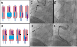
The technical considerations for the above methods, including specific techniques such as distal balloon deflection and distal balloon deflation techniques (Figure 3) are illustrated in detail in the online version of this chapter of Tools & Techniques.

Figure 3. Distal balloon deflation technique. (A) Possible mechanism: the inflation of distal balloon may give the space to cross the proximal stent. (B) Possible mechanism: the inflation of distal balloon may change the route of the proximal stent. (C) RCA CTO lesion (D,E) Just after the deflation of distal balloon, proximal stent was able to be delivered to the lesion location. (F) Final result.

