Tako-Tsubo syndrome is a transient cardiomyopathy that mimics acute coronary syndrome in the absence of obstructive coronary artery disease characterised by chest pain, ST-segment alteration, minimal increase of cardiac enzymes and balloon-like asynergy of the apical region, frequently triggered by a physical or mental stress preceding the symptoms onset.
In the case reported, the syndrome was observed in a 61-year-old woman with chest pain, ST-segment elevation and negative T-waves in anterior ECG leads V1 - V4 as well as a minimal increase in troponin I and creatine kinase-MB. The patient was referred to our catheterisation laboratory with a supposed diagnosis of STEMI. Coronary angiography showed a diffuse spasm of the anterior interventricular artery (IVA) below the first septal branch (Video 1; Figure 1a).
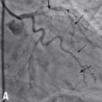
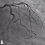
Figure 1. Diffuse spasm (arrows) of the IVA below the first septal branch (a); IVA after verapamil bolus (0.2 mg) (b).
The spasm was resolved by intracoronary verapamil (0.2 mg) (Video 2; Figure 1b). Intravascular ultrasound excluded the presence of atherosclerotic plaques. Left ventriculography showed an apical ballooning with hyper-contractility of the basal segments and an ejection fraction (EF) of 36% (Video 3; Figure 2a). Nine days later, left ventricular contractility was normalised with an EF of 54% (Video 4; Figure 2b).
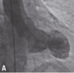
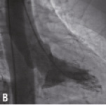
Figure 2. Left ventriculography at presentation showing apical ballooning (a); left ventriculography nine days after the acute phase showing normal left ventricular motion (b).
The pathophysiology of the syndrome is controversial and several hypothesises are considered, including catecholamine-mediated myocardial stunning and endothelial dysfunction. Although there are contrasting opinions about the role played by coronary spasm, we believe that its angiographic demonstration in the acute phase may represent an evidence that this mechanism can be one of the triggers precipitating the syndrome.
Online data supplement
Video 1. Diffuse spasm of the IVA below the first septal branch.
Video 2. IVA after verapamil infusion.
Video 3. Left ventriculography at presentation showing apical ballooning.
Video 4. Left ventriculography nine days after the acute phase showing normal left ventricular motion.
Supplementary data
To read the full content of this article, please download the PDF.
Moving image 1.
Moving image 2.
Moving image 3.
Moving image 4.
