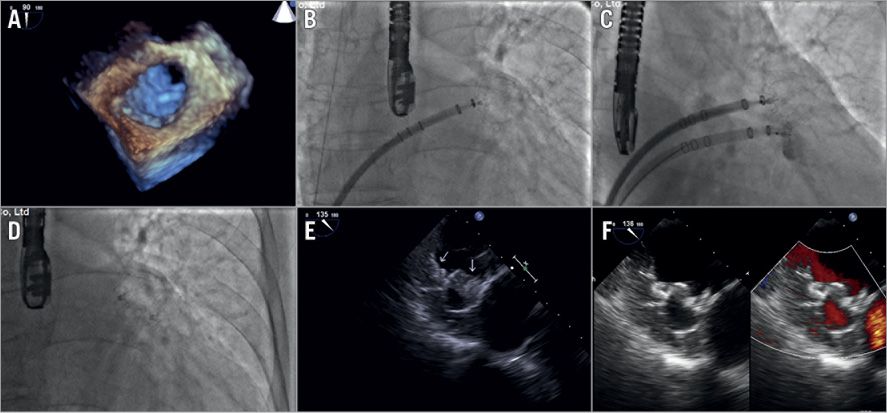

Figure 1. Angiography and TOE imaging of the kissing-Watchman procedure and follow-up. A) Three-dimensional TOE revealed the large LAA ostium. B) The 27 mm WATCHMAN device was pre-released and held still at the intended landing zone. C) A second WATCHMAN device was placed adjacent to the 27 mm device. D) The two WATCHMAN devices in a kissing fashion. E) TOE at three-month follow-up showed adequate LAA sealing, and no device-related thrombus was identified. F) TOE at one-year follow-up showed adequate LAA sealing, without device-related thrombus.
The WATCHMAN™ device (Boston Scientific, Marlborough, MA, USA) is the predominant occluder used in China because of medical insurance policies. Though there are larger sizes in other devices nowadays, the maximum commercially available WATCHMAN device is 33 mm, which only fits a left atrial appendage (LAA) with a maximum diameter of 30 mm. In patients with multilobed LAA and a large LAA ostium, the implantation of two devices, either in a one-stop or in a staged procedure to seal the LAA completely, is feasible as reported by previous studies1-4. However, it is unknown whether this approach is feasible in the setting of a single lobe with a giant ostium (>30 mm). We describe here the simultaneous kissing-Watchman technique to occlude a single lobe LAA with large ostium anatomy.
The patient was a 70-year-old male with atrial fibrillation (CHA2DS2-VASc=3) on warfarin who was referred for LAA closure due to gastrointestinal bleeding. Transoesophageal echocardiography (TOE) showed a single lobe LAA with a maximum LAA ostium of 35 mm (Moving image 1). Three-dimensional TOE confirmed the large LAA ostium (Figure 1A). Therefore, use of the kissing-Watchman technique was planned, with two WATCHMAN devices in 27 mm and 21 mm sizes. The 27 mm WATCHMAN device was pre-released and held still at the intended implantation zone (Figure 1B, Moving image 2). A second transseptal puncture was performed, and the 21 mm WATCHMAN device was placed carefully next to the 27 mm device in a kissing fashion (Figure 1C, Moving image 2). The tug test was performed on the two devices by pulling the parallel delivery systems simultaneously (Moving image 2). The two devices were released one after the other when the PASS criteria had been met (Figure 1D, Moving image 2). TOE at three-month and one-year follow-up showed adequate LAA sealing, and no device-related thrombus was identified (Figure 1E, Figure 1F, Moving image 3). Full-dose rivaroxaban was prescribed for the first three months post operation, then dual antiplatelet therapy for the next eight months. Clinical observation during the 12-month follow-up revealed no severe complications or major adverse events.
Conflict of interest statement
The authors have no conflicts of interest to declare.
Supplementary data
To read the full content of this article, please download the PDF.
Moving image 1. TOE imaging of the single lobe LAA with large ostium.
Moving image 2. Angiography imaging of the kissing-Watchman procedure.
Moving image 3. TOE follow-up.

