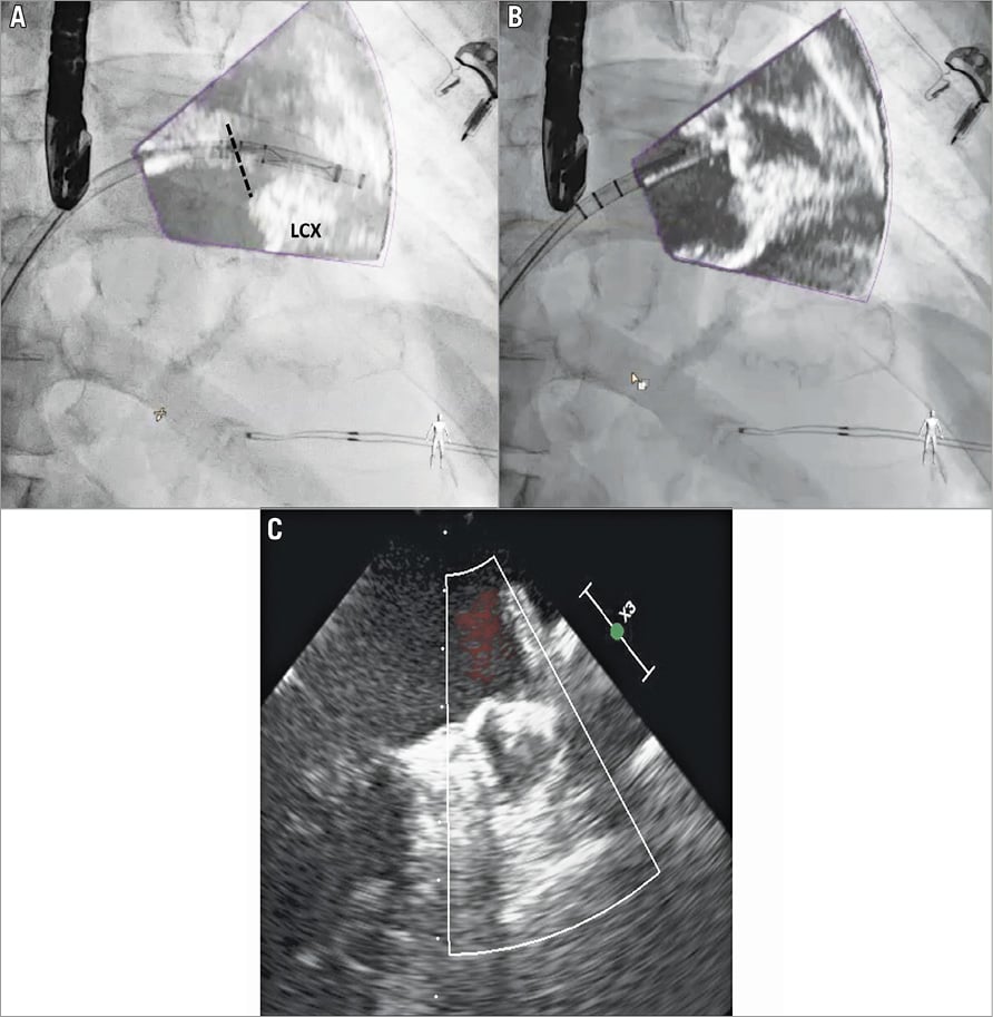

An 84-year-old man with paroxysmal atrial fibrillation (AF) was scheduled for percutaneous left atrial appendage (LAA) closure. The patient had a high thromboembolic risk based on a CHA2DS2-VASc score of 5 (previous stroke, age >75 years and hypertension) and a prior history of ulcerative colitis with recurrent episodes of major gastrointestinal bleeding under oral anticoagulant therapy. Due to the presence of severe chronic kidney disease (estimated glomerular filtration rate [eGFR] 28 ml/min/1.73 m2), an attempt was made to perform the procedure without contrast injection using the EchoNavigator system (Philips Healthcare, Best, the Netherlands), which allows real-time fusion of 3D transoesophageal echocardiography (TEE) and fluoroscopic images1. Following transseptal puncture, adequate LA filling pressure (14 mmHg) was confirmed and procedural 3D TEE measurements showed a maximal and mean diameter of the landing zone of 25.3 mm and 23.5 mm, respectively. It was decided to implant a 27 mm WATCHMAN™ device (Boston Scientific, Marlborough, MA, USA). The WATCHMAN access sheath was advanced over a 5 Fr pigtail catheter into the LAA cavity. Thanks to the projection of TEE images on the fluoroscopic screen (“fused image”), we could precisely position the 27 mm marker of the access sheath at the level of the landing zone without contrast injection (Panel A, black dotted line). The WATCHMAN device was then released by careful retraction of the access sheath and the correct final position was confirmed by both standard TEE and fused images, without peri-device leak (Panel B, Panel C, Moving image 1). The total procedure duration was 45 minutes.
Prevalence of chronic kidney disease is high in patients undergoing LAA occlusion, approaching 40%2. This case illustrates a potential role of fusing echocardiography and fluoroscopy images during LAA closure with the WATCHMAN device in patients with advanced renal failure in whom contrast injection may be contraindicated. Since the EchoNavigator system relies on optimal 3D-TEE visualisation of the LAA, difficult image acquisition or poor visualisation of the LAA distal lobes during sheath placement may represent a potential technical limitation of this approach.
Conflict of interest statement
A. Aminian is consultant and proctor for Boston Scientific. The other authors have no conflicts of interest to declare.
Supplementary data
Moving image 1. TEE and X-ray fusion during WATCHMAN implantation.
Supplementary data
To read the full content of this article, please download the PDF.
Moving image 1. TEE and X-ray fusion during WATCHMAN implantation.

