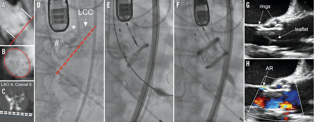
This case report demonstrates the potential for contrast agent-free preprocedural planning (Figure 1A-C) and transcatheter aortic valve implantation (TAVI; Figure 1D-H) of a Direct Flow Medical® (DFM) transcatheter aortic valve (Direct Flow Medical, Santa Rosa, CA, USA), a device without a metallic frame, in an 84-year-old patient with renal failure and a severe aortic valve stenosis. Avoiding any application of contrast agent, and therefore its related complications, might extend even further the indication for TAVI implantation in such high-risk patients. This approach might currently be limited to certain anatomies and special clinical situations until larger studies confirm the strategy.

Figure 1. Pre- and intraprocedural imaging. Using non-contrast 3D FLASH MRA and no-contrast CT (A), the distance to the right/left coronary ostium was measured perpendicular to the annular plane. B) The cross-sectional area was assessed by means of planimetry, and (C) an optimal fluoroscopic angulation for the DFM implantation was assessed with all three leaflets in line. Pigtail catheters were placed in the NCC (#) and RCC (*) to draw an imaginary line for identification of the landing zone for the ventricular DFM ring (D). Positioning (E) and final fluoroscopic result (F) of the DFM implantation with an optimal position. TEE confirms optimal position of the DFM valve with only trace paravalvular AR (G & H).
Conflict of interest statement
J. Reinöhl is a proctor for Direct Flow Medical. F. De Marco and A. Latib are consultants for Direct Flow Medical. The other authors have no conflicts of interest to declare.

