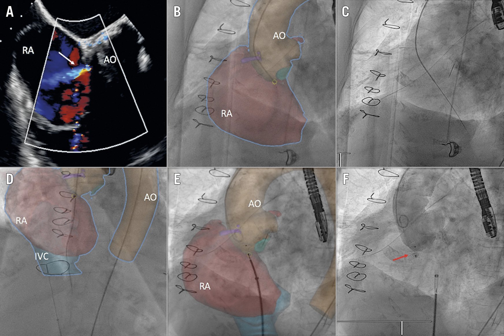
Figure 1. Cardiac fusion imaging for procedural guidance. A) 2D TEE Doppler colour image showing the aorto-right atrial shunt (white arrow). B) CTA-fluoro fusion image: cardiac fusion imaging guidance to cross the fistula from the aorta (AO) to the right atrium (RA). C) Fluoro image: stiff wire crossing from the aorta into the right atrium. D) CTA-fluoro fusion image: wire advanced and snared into the inferior vena cava (IVC). E) CTA-fluoro fusion image: device deployment guided by cardiac fusion imaging. F) Fluoro image: final result showing the AMPLATZER Duct Occluder (ADO) device (red arrow) sealing the aorto-atrial fistula.
A 75-year-old patient with a previous history of heart valve surgery was admitted due to recurrent congestive heart failure.
In 1993 she had undergone open heart surgery where the aortic valve was replaced with a mechanical prosthesis. In 2015, an ascending aortic aneurysm replacement was performed with a Dacron graft including the non-coronary sinus.
During a congestive heart failure episode in 2017, an aorto-right atrial shunt was observed on transthoracic echocardiography. The patient was not considered for cardiac surgery because she was too high risk.
After a new heart failure episode, the patient was referred to our centre for evaluation of possible percutaneous closure of the aorto-atrial fistula. Transoesophageal echocardiography (TEE) confirmed a 5 mm high-flow fistula between the non-coronary sinus and the right atrium (Figure 1A).
The procedure was performed under general anaesthesia guided by fluoroscopy, TEE and computed tomography angiography (CTA)-fluoroscopy fusion imaging. Vascular access was gained via the right femoral vein and left femoral artery. Initial aortography with a pigtail catheter showed the aorto-atrial fistula (Moving image 1). Then, with cardiac fusion imaging guidance and landmarks provided by angiography and tissue calcification, the fistula was easily crossed using a Terumo stiff wire (Terumo Corp., Tokyo, Japan) with a multipurpose catheter (Figure 1B, Figure 1C).
After that, the wire was advanced and snared into the inferior vena cava (Figure 1D) and externalised via a right femoral vein sheath creating an arteriovenous loop.
Finally, the delivery sheath was advanced from the venous side and the 8/6 mm AMPLATZER™ Duct Occluder (ADO; Abbott Vascular, Santa Clara, CA, USA) deployed (Figure 1E, Moving image 2), achieving complete sealing of the fistula (Figure 1F, Moving image 3).
The patient had an uneventful recovery and remains asymptomatic after one-year follow-up.
Conflict of interest statement
I. Cruz-Gonzalez is a proctor for Abbott Vascular. The other authors have no conflicts of interest to declare.
Supplementary data
To read the full content of this article, please download the PDF.
Moving image 1. Aortography showing the aorto-atrial fistula.
Moving image 2. Device deployment guided by cardiac fusion imaging.
Moving image 3. Aortography showing the ADO device sealing the fistula completely.