IMAGE IN CARDIOLOGY

DOI: 10.4244/EIJV12I10A215

Overstepping anatomical hurdles when opening an extremely rudimentary right ventricular outflow tract - greater safety via a hybrid strategy?

Rouven Kubicki1, MD; Johannes Kroll2, MD; Brigitte Stiller1, MD, PhD; Jochen Grohmann1*, MD

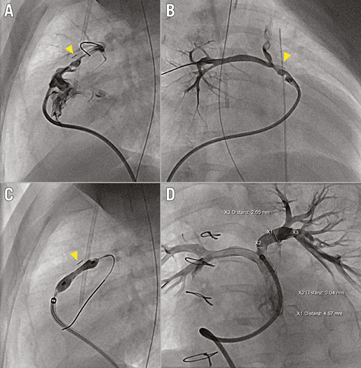
Managing pulmonary atresia with ventricular septal defect and major aortopulmonary collaterals (PA/VSD/MAPCAs) is challenging. Strategies for rehabilitating diminutive native pulmonary arteries may be considered. Right ventricular outflow tract (RVOT) stenting is a promising approach in this situation. We hereby introduce a modified hybrid approach we recently undertook in a neonate weighing 3.31 kg. First, a midline sternotomy was performed, followed by placing a pericardial safety cuff around the native main pulmonary artery (MPA), and a vascular clip serving as a landmark for bifurcation. The chest was left open to permit quick and controlled access to any bleeding site in case of accidental perforation. After transfer to the cathlab, the atretic RVOT tunnel was opened via radiofrequency perforation and a coronary wire advanced to the distal right pulmonary artery, followed by balloon dilation. Selective angiography showed the rudimentary infundibulum and MPA (Panel A, Moving image 1) as well as the diminutive branch pulmonary arteries (Panel B, Moving image 2). RVOT-to-MPA stenting was facilitated by the marking clip (∆) proximal to the bifurcation (Panel C, Moving image 3). Finally, the chest was closed in the operating room. Eight weeks later, follow-up angiography confirmed catch-up growth of the native pulmonary arteries induced by antegrade flow (Panel D, Moving image 4).

Conflict of interest statement

The authors have no conflicts of interest to declare.

Supplementary data

Moving image 1. Angiography infundibulum, lateral view.

Moving image 2. Angiography MPA, RAO.

Moving image 3. Stenting RVOT to MPA, lateral view.

Moving image 4. Follow-up result after eight weeks, LAO.

Supplementary data

To read the full content of this article, please download the PDF.

Angiography infundibulum, lateral view.

Angiography MPA, RAO.

Stenting RVOT to MPA, lateral view.

Follow-up result after eight weeks, LAO.
Volume 12 Number 10
Nov 20, 2016
Volume 12 Number 10
View full issue


Key metrics

Suggested by Cory

Image – Interventional flashlight

10.4244/EIJ-D-19-00962 Sep 18, 2020
Melody valve implantation in tetralogy of Fallot with acquired double right ventricular outflow tract
Valderrama P et al
free

EXPERT REVIEW

10.4244/EIJ-D-17-00501 Sep 24, 2017
Percutaneous pulmonary valve implantation: what have we learned over the years?
Boudjemline Y
free

Image – Interventional flashlight

10.4244/EIJ-D-19-00649 Oct 9, 2020
Fistula between the right pulmonary artery and left atrium in a newborn: management and successful interventional treatment
Michalak K et al
free

Clinical research

10.4244/EIJ-D-18-00857 Feb 5, 2021
Long-term follow-up after recanalisation of aortic arch atresia
Chessa M et al
free
Trending articles
152.9

Clinical research

10.4244/EIJ-D-20-01125 Oct 20, 2021
An upfront combined strategy for endovascular haemostasis in transfemoral transcatheter aortic valve implantation
Costa G et al
free
47.8

NEW INNOVATION

10.4244/EIJ-D-15-00467 Feb 20, 2018
Design and principle of operation of the HeartMate PHP (percutaneous heart pump)
Van Mieghem NM et al
free
39.1

Clinical research

10.4244/EIJ-D-22-00558 Feb 6, 2023
Permanent pacemaker implantation and left bundle branch block with self-expanding valves – a SCOPE 2 subanalysis
Pellegrini C et al
free
X

The Official Journal of EuroPCR and the European Association of Percutaneous Cardiovascular Interventions (EAPCI)

EuroPCR EAPCI
PCR ESC
Impact factor: 7.6
2023 Journal Citation Reports®
Science Edition (Clarivate Analytics, 2024)
Online ISSN 1969-6213 - Print ISSN 1774-024X
© 2005-2024 Europa Group - All rights reserved