IMAGE IN CARDIOLOGY

DOI: 10.4244/EIJV11I7A162

Neonatal tricuspid stenosis caused by device closure of a large coronary fistula

Chingying Changchien, MD; Ming-Tai Lin*, MD, PhD; Ching-Chia Wang, MD, PhD; Hsin-Ming Liu, MD; Chia-Ching Wang, MD; Shuenn-Nan Chiu, MD, PhD; Mei-Hwan Wu, MD, PhD; Jou-Kou Wang, MD, PhD

A male baby, weighing 3,476 gm, was noted to have grade III/VI continuous murmur. Cardiomegaly was noted on his chest x-ray (Figure 1A). Echocardiography (Figure 1B) showed a large right coronary arteriovenous fistula (CAVF) with drainage to the right ventricle. Computed tomography (Figure 1C) demonstrated similar findings. Transcatheter closure of the 3.5 mm fistula was attempted at six days of age. Coronary cineangiography confirmed the diagnosis (Figure 1D, Moving image 1). An Amplatzer™ Duct Occluder II (9-PDA2-04-04; St. Jude Medical, St. Paul, MN, USA) was deployed smoothly (Figure 1E and 1H, Moving image 2). The post-procedural course was uneventful except for fluctuating saturation (90-95%). Echocardiography showed an acceleration of tricuspid antegrade flow (Figure 1F, Moving image 3), which may be related to the compression on the right atrioventricular groove by the device: this caused right to left shunt at the level of the foramen ovale. His saturation normalised four months later and echocardiography revealed the resolution of tricuspid stenosis. Concurrent computed tomography (Figure 1G) also showed that the fistula was completely occluded.
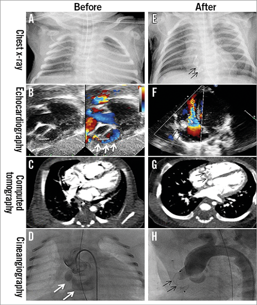
Figure 1. Image studies before and after transcatheter closure of a coronary fistula. (A-D) Before the closure, (E-H) after the closure, (F) device-related tricuspid stenosis. White arrows: coronary arteriovenous fistula; dark arrows: occluder

We suggest that the Amplatzer Duct Occluder II might be feasible for transcatheter closure of neonatal CAVF. Transient cyanosis due to device-related tricuspid stenosis should be monitored.

Conflict of interest statement

The authors have no conflicts of interest to declare.

Supplementary data

Moving image 1. Selective cineangiogram, lateral view of right coronary arteries.

Moving image 2. Ductal occluder deployment process.

Moving image 3. Tricuspid stenosis.
Supplementary data

To read the full content of this article, please download the PDF.

Moving image 1. Selective cineangiogram, lateral view of right coronary arteries.

Moving image 2. Ductal occluder deployment process.

Moving image 3. Tricuspid stenosis.

Volume 11 Number 7
Nov 20, 2015
Volume 11 Number 7
View full issue


Key metrics

Suggested by Cory

Image – Interventional flashlight

10.4244/EIJ-D-19-00649 Oct 9, 2020
Fistula between the right pulmonary artery and left atrium in a newborn: management and successful interventional treatment
Michalak K et al
free

10.4244/EIJV12SXA6 May 16, 2016
Coronary-cameral fistulas: indications and methods for closure
Carminati M et al
free

IMAGE IN CARDIOLOGY

10.4244/EIJV12I8A164 Oct 10, 2016
Closure of a coronary artery: coronary sinus fistula
George S et al
free

IMAGE IN CARDIOLOGY

10.4244/EIJV12I8A165 Oct 10, 2016
Percutaneous closure of an iatrogenic aortocoronary venous fistula after coronary bypass graft surgery
Sandhu K et al
free

CLINICAL RESEARCH

10.4244/EIJ-D-17-00632 Apr 6, 2018
Combined use of coils and Onyx for transcatheter closure of coronary artery fistulae
Li P et al
free

IMAGE IN CARDIOLOGY

10.4244/EIJV12I9A193 Oct 20, 2016
Large left lung mass: an unexpected finding
Henzel J et al
free

CLINICAL RESEARCH

10.4244/EIJ-D-15-00238 Apr 20, 2017
Are the AMPLATZER Duct Occluder II Additional Sizes devices dedicated only for smaller children?
Fiszer R et al
free

Image – Interventional flashlight

10.4244/EIJ-D-19-00198 Jun 12, 2020
Percutaneous closure of atrial fistula after ascending aortic aneurysm surgery using cardiac fusion imaging
Cruz-González I et al
free
Trending articles
309.93

State-of-the-Art Review

10.4244/EIJ-D-21-00695 Nov 19, 2021
Transcatheter treatment for tricuspid valve disease
Praz F et al
free
166.7

Expert review

10.4244/EIJ-D-21-00690 May 15, 2022
Crush techniques for percutaneous coronary intervention of bifurcation lesions
Moroni F et al
free
92.45

State-of-the-Art Review

10.4244/EIJ-D-20-01296 Aug 27, 2021
Management of cardiogenic shock
Thiele H et al
free
72.85

State-of-the-Art

10.4244/EIJ-D-24-00386 Feb 3, 2025
Mechanical circulatory support for complex, high-risk percutaneous coronary intervention
Ferro E et al
free
58.3

Clinical research

10.4244/EIJ-D-23-00344 Sep 18, 2023
Clinical outcomes of TAVI with the Myval balloon-expandable valve for non-calcified aortic regurgitation
Sanchez-Luna JP et al
free
56.65

Clinical research

10.4244/EIJ-D-20-01155 Oct 20, 2021
A deep learning algorithm for detecting acute myocardial infarction
Liu W et al
free
33.9

CLINICAL RESEARCH

10.4244/EIJ-D-17-00381 Oct 11, 2017
Stent malapposition and the risk of stent thrombosis: mechanistic insights from an in vitro model
Foin N et al
free
33.65

State-of-the-Art

10.4244/EIJ-D-23-00606 Jan 1, 2024
Targeting inflammation in atherosclerosis: overview, strategy and directions
Waksman R et al
free
26

Expert Review

10.4244/EIJ-D-24-00535 May 5, 2025
Catheter-based techniques for pulmonary embolism treatment
Costa F et al
X

The Official Journal of EuroPCR and the European Association of Percutaneous Cardiovascular Interventions (EAPCI)

EuroPCR EAPCI
PCR ESC
Impact factor: 7.6
2023 Journal Citation Reports®
Science Edition (Clarivate Analytics, 2024)
Online ISSN 1969-6213 - Print ISSN 1774-024X
© 2005-2025 Europa Group - All rights reserved