The treatment of simple congenital malformations has evolved significantly over the last decade. Lesions such as septal defects, patent ductus arteriosus (PDA), coarctation, conduit dysfunction, pulmonary artery stenosis, and fistulae are now all addressed via a transcatheter approach as first-line treatment. The field of structural interventions is a spin-off of interventional congenital cardiology. The first successful percutaneous valve was the transcatheter pulmonary valve. Procedures such as paravalvular leak closure and post-myocardial infarction ventricular septal rupture closure are all extrapolations of interventional congenital procedures. Furthermore, imaging which is essential in congenital cardiology for understanding complex malformations, has now spilled over into the structural arena for understanding and planning procedures.
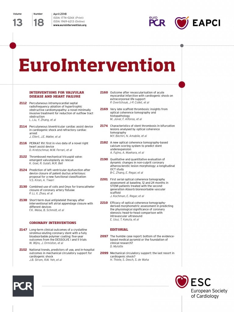
In this issue of EuroIntervention, two articles examine the treatment of congenital vascular connections. The first article, entitled “Combined use of coils and Onyx for transcatheter closure of coronary artery fistulae”1 written by Li and colleagues, examines the safety and efficacy of coronary artery fistula (CAF) closure using the combination of coils and Onyx® Liquid Embolic System (Micro Therapeutics, Inc., Irvine, CA, USA), a novel approach.
The second article, entitled “Prediction of left ventricular dysfunction after device closure of patent ductus arteriosus: proposal for a new functional classification”2 written by Kiran and Tiwari, examines the association between PDA size and left ventricular dysfunction after closure using traditional PDA closure devices.
Both articles examine the safety, efficacy, and complications associated with fistula closure.
Coronary artery fistulae are congenital malformations of the coronary arteries. CAF occur when a coronary artery vessel communicates directly with a low-pressure right-sided structure without an intervening capillary network3,4. CAF closure can be achieved by either the surgical or the transcatheter route. When technically feasible, the transcatheter approach is the preferred route given its less invasive nature. Devices used successfully to close CAF include coils, umbrella devices, covered stents, and the AMPLATZER family of devices. By far the most common device used worldwide is the coil5.
As with any procedure, there are risks. The most feared short-term complication of CAF closure is myocardium necrosis resulting from reflux into the native coronary tree. The long-term complication is recanalisation of the CAF. Coil closure of CAF is generally safe; however, the recanalisation rates are high, especially with large fistulae. Also, effective closure usually requires the use of many coils which adds to the cost of the procedure. More recently, the use of other devices such as the vascular plug has improved the short- and long-term efficacy of CAF closure without increasing the risk6.
The article published by Li and colleagues is a retrospective, single-centre study1. It enrolled 26 consecutive patients for CAF closure using the combination of coils and Onyx. The majority of the CAF connected to the pulmonary artery, and all patients were symptomatic.
The procedure involved first the placement of detachable coils in the fistula followed by the direct injection of Onyx into the fistula. The initial deployment of coils served first to reduce coronary flow significantly, and second to act as a scaffold for the injection of Onyx. Onyx is a liquid embolic system used mostly in the neurological field. It is a mixture of ethylene-vinyl alcohol copolymer (EVOH), dissolved in dimethyl-sulfoxide (DMSO) and mixed with micronised tantalum powder for radiopaque visualisation. Onyx will precipitate immediately when it comes into contact with water, blood, or saline. DMSO keeps the Onyx soluble.
Onyx is injected into the fistula using a microcatheter the lumen of which is filled with DMSO to prevent precipitation in the catheter. DMSO, if released from the microcatheter too quickly, can induce vasospasm and angionecrosis. The preparation and application of Onyx requires a particular step-by-step application which, if it is not followed carefully, will lead to angiotoxicity, increased reflux and catheter entrapment7.
Immediate fistula occlusion was achieved in 88.5% of patients. Assessment of recanalisation was performed at a median of 11.5 months. Selective coronary angiography was performed in 57% of patients, and the remaining were assessed by CT angiography. At 11.5 months 80.8% were still occluded. The only notable complication was a significant rise in cardiac troponin I and creatinine kinase-MB levels 24 hours post procedure. However, the levels returned to baseline at 30 days.
The primary purpose of the study was to assess the safety and both the immediate and long-term efficacy of CAF embolisation using the combination of coils and Onyx. Li and colleagues are to be congratulated. They have demonstrated a novel approach to the transcatheter treatment of CAF. There are, however, three issues of concern.
First, from a safety perspective, whenever a non-retrievable device/product is used for CAF occlusion there is a concern of reflux and embolisation into the native coronary vasculature causing myocardial necrosis. The study describes a significant increase in troponin levels 24 hours after the procedure that normalised one month later. No explanation is given for this effect, and no assessment of left ventricular function is provided. The rise in cardiac enzymes is small, and perhaps it is related to catheter manipulation within the coronary tree, but it does raise the concern that a small amount of product may have refluxed into the native coronary circulation.
Second, the preparation of Onyx is cumbersome. Onyx is used mostly in the interventional neurological field for the embolisation of arteriovenous malformations of the brain and spinal cord. When used carefully in experienced hands it is reported to have rare complications and effective closure rates. However, its laborious preparation and unique risk profile may limit its generalisability.
Lastly, the assessment of recanalisation may be faulty. The most common long-term complication of CAF embolisation is recanalisation. The recanalisation rate at 11.5 months in this study was only 19%. However, only 57% of patients were assessed using direct coronary angiography; the remainder were assessed by CT angiography. CT angiography may underestimate recanalisation rates compared to direct coronary injection since direct coronary angiography allows selective injection at higher pressures. Therefore, the recanalisation rates may be underestimated.
Overall, Li and colleagues have demonstrated that the use of coils plus Onyx in experienced hands is safe, effective, and less costly than coils alone for the treatment of CAF. The laborious preparation of Onyx and the potential for uncontrolled reflux may hinder its acceptance in the interventional cardiology community. Eventually, comparisons with other approaches will have to be made to determine which is superior. Regardless, the study offers another effective strategy for CAF treatment. Li and colleagues have provided the interventional cardiology community with another tool for the transcatheter treatment of CAF.
Occlusion of the patent ductus arteriosus is one of the most common structural interventional procedures and can be carried out safely and effectively using either a device or a coil8. In this single-centre, retrospective, cross-sectional analysis of 447 patients undergoing device closure of a patent ductus arteriosus (PDA), Kiran and Tiwari propose a new stratification based on the risk of developing left ventricular (LV) dysfunction in the days following the intervention2. The cohort was arbitrarily divided into four groups according to the angiographic measurement of the PDA at its pulmonary end indexed to the body surface. All patients had close clinical and echocardiographic monitoring in the immediate post-intervention period and treatment was promptly initiated according to the European Heart Failure Guidelines.
The study suggests that the risk of echocardiographic LV systolic dysfunction increases proportionally to the indexed PDA size and is frequent for values superior to 6 mm/m2. Consequently, the authors propose that the indexed PDA size is a good predictor of LV dysfunction post occlusion and could be used to anticipate the level of surveillance needed.
As indicated by the authors, sudden elimination of a large ductal left to right shunt produces an acute increase in LV afterload by removal of a low-pressure resistor and a concomitant decrease in preload, further challenging the left ventricle on the Frank-Starling curve. Systolic dysfunction of the left ventricle has been described after PDA surgical ligation or percutaneous embolisation9 but usually resolves after a period of time, especially when the ejection fraction was normal before the intervention. The myocardium adapts to the new physiological state and gradually remodels towards normal function.
In our experience, symptomatic heart failure in the immediate post-occlusion period is exceptionally uncommon and rarely requires medical intervention. This study focuses solely on echocardiographic indices of LV dysfunction to ascertain haemodynamic impact post PDA occlusion. This is problematic, as the heart failure severity is usually graded according to recognised clinical criteria that include more than just echocardiographic parameters10,11. Moreover, the pertinence of the quoted predominantly adult heart failure guidelines12 to direct management in an otherwise healthy but volume-loaded myocardium is questionable. With this in mind, the applicability of the proposed invasive post-occlusion surveillance and treatment protocol is unclear within the accepted practice framework that exists in developed countries.
This study proposes a simple and intuitive risk stratification of patients undergoing percutaneous duct occlusion based on the size of the structure indexed to body surface area. The findings of this study are particularly relevant to healthcare systems in which resources are limited, and clinical vigilance needs to be prioritised to high-risk patients. Although this paper raises awareness about the high incidence of post-occlusion left ventricular dysfunction for large PDAs, its applicability to current practice in developed countries is unclear.
Conflict of interest statement
The authors have no conflicts of interest to declare.

