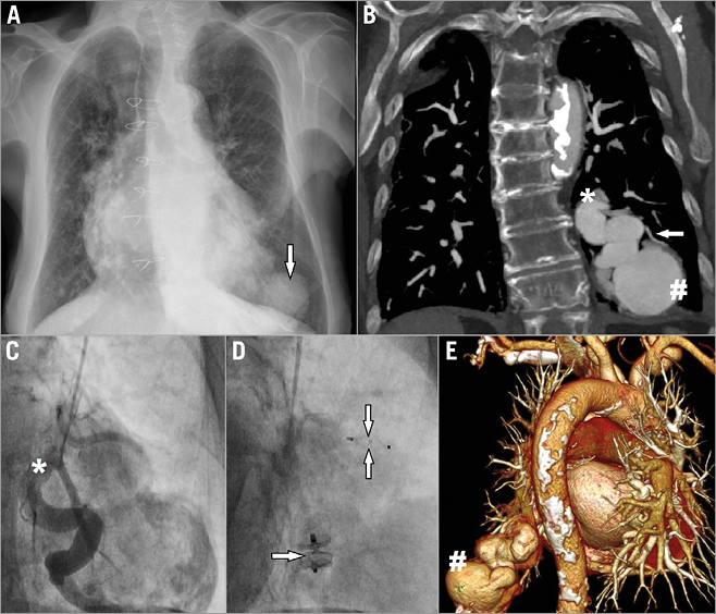
An 83-year-old female with a history of two coronary artery bypass graft surgeries and multiple comorbidities presented with aggravation of chronic heart failure accompanied by central cyanosis and nail clubbing. Pulse oximetry was 80-85% on room air, 95% on supplemental oxygen. Chest X-ray showed a large mass in the posterobasal area of the left lung (Panel A, arrow). Common states such as pneumonia or lung cancer were initially suspected, yet, surprisingly, contrast-enhanced computed tomography revealed a giant, 52 mm pulmonary arteriovenous fistula (Panel B, Panel E, #). The structure derived from the segmental branch of the left pulmonary artery (Panel B, Panel C, asterisk); an additional feeding artery was also identified (Panel B, arrow). Both vessels were closed percutaneously using two AMPLATZER™ devices (AVP II 10 mm and AVP IV 4 mm, St. Jude Medical, St. Paul, MN, USA) (Panel D, single and double arrow) with good clinical outcome; oxygen saturation increased up to 98%.
Pulmonary arteriovenous fistula (PAVF) is a direct communication between the branches of pulmonary arteries and pulmonary veins, often presenting with aneurysmal enlargement of the feeding vessel. PAVFs are usually hereditary and mostly associated with haemorrhagic telangiectasia, also known as Osler-Weber-Rendu syndrome. Acquired, post-traumatic (including iatrogenic) forms have also been described. Thin and fragile walls make a PAVF prone to rupture, which may lead to dangerous pulmonary haemorrhage. Other possible complications include neurological events (stroke or cerebral abscess caused by paradoxical embolism), migraines and hypoxaemia.
Regardless of symptoms and aetiology, a fistula with the feeding artery of >3 mm in diameter should be qualified for treatment. When technically feasible, percutaneous closure is currently preferred over surgical treatment.
Conflict of interest statement
The authors have no conflicts of interest to declare.
Supplementary data
Moving image 1. Baseline selective angiography of the feeding vessel.
Moving image 2. Selective angiography of the feeding vessel directly after the procedure.
Supplementary data
To read the full content of this article, please download the PDF.
Baseline selective angiography of the feeding vessel.
Selective angiography of the feeding vessel directly after the procedure.