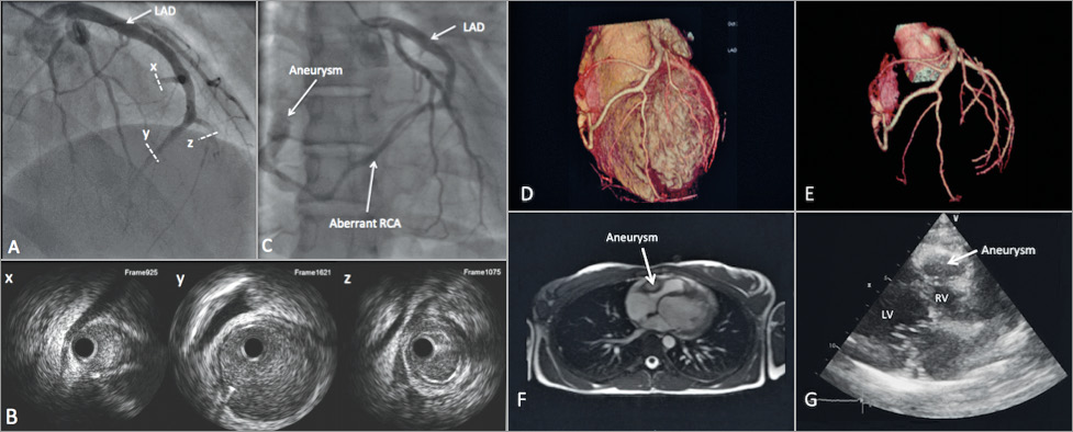
A 25-year-old female patient without a past medical history was admitted for acute onset of retrosternal chest pain with an electrocardiogram displaying low QRS voltage and dominant apical Q-wave with troponin elevation. Coronary angiography showed a single coronary artery (SCA) arising from the left aortic sinus and an abnormal right coronary artery (RCA) originating from the left anterior descending (LAD) artery with multiple dissections (distal LAD artery, the RCA and an accessory branch) confirmed using intravascular ultrasound imaging (Panel A, Panel B, Moving image 1). We adopted a conservative treatment due to withdrawal of symptoms. A control coronary angiography on day seven showed recanalisation of the dissected branches. The RCA terminated at the lateral side of the right ventricle in a polylobulated coronary artery aneurysm (CAA) (Panel C, Moving image 2). Contrast-enhanced computed tomography confirmed the absence of the RCA ostium and showed a nodular formation (34×17×32 mm) exerting a mass effect on the roof of the right ventricle (Panel D, Panel E). Magnetic resonance imaging showed an ovoid formation with heterogeneous contrast enhancement corresponding to the CAA (Panel F). Echocardiography showed the aneurysm on the lateral part of the right heart beneath the insertion of the tricuspid valve with no haemodynamic effect on the RV (Panel G, Moving image 3). Six months later, the size of the aneurysm was stable and the clinical course was uneventful. To the best of our knowledge, this is the first case combining a triple association of an SCA, spontaneous multiple intimal dissections and a CAA in a young woman with acute coronary syndrome.
Conflict of interest statement
The authors have no conflicts of interest to declare.
Supplementary data
Moving image 1. Coronary angiography at admission (left anterior oblique cranial view) showing multiple intimal dissections of the distal LAD with fresh thrombus, the RCA and an accessory aberrant branch (above the birth of the RCA).
Moving image 2. Rotational coronary angiography on day seven showing recanalisation of the distal LAD and the RCA. The RCA terminates at the lateral side of the right ventricle in a polylobulated aneurysm.
Moving image 3. Echocardiography showing a nodular formation located on the lateral part of the right heart beneath the insertion of the tricuspid valve.
Supplementary data
To read the full content of this article, please download the PDF.
Coronary angiography at admission (left anterior oblique cranial view) showing multiple intimal dissections of the distal LAD with fresh thrombus, the RCA and an accessory aberrant branch (above the birth of the RCA).
Rotational coronary angiography on day seven showing recanalisation of the distal LAD and the RCA. The RCA terminates at the lateral side of the right ventricle in a polylobulated aneurysm.
Echocardiography showing a nodular formation located on the lateral part of the right heart beneath the insertion of the tricuspid valve.