A 48-year-old woman with previous history of hyperthyroidism caused by Graves' disease presented with unstable angina. Three years before, the patient was admitted for a non-ST-segment elevation myocardial infarction. At that time, coronary angiography revealed severe coronary artery tortuosity with an abrupt and diffuse lumen narrowing of the distal left anterior descending coronary artery (LAD) as a result of spontaneous coronary artery dissection (SCAD) presenting as an intramural haematoma (Figure 1A, Moving image 1) with no stenosis in the right coronary artery (RCA) (Figure 1B, Moving image 2). The patient was successfully managed conservatively, and further angiography screening identified stigmas of fibromuscular dysplasia in the right renal artery (Figure 1E).
The current coronary angiogram showed complete healing (restitutio ad integrum) of the distal LAD and a mild stenosis with a small aneurysm (not present in the previous angiogram) in the proximal RCA (Figure 1C, Figure 1D, Moving image 3, Moving image 4). Of note, no classic angiographic findings of SCAD were observed. Nevertheless, along with the characteristic double-lumen pattern (Figure 1F, Figure 1G), optical coherence tomography (OCT) disclosed a “lotus root” appearance with multiple thick intimal septa splitting the false lumen into small, rounded channels in the proximal RCA. Multiple circular concave-edged channels caused by smooth-edged, signal-rich septa, were visualised (Figure 1H, Figure 1I, Moving image 5).
SCAD is a relatively uncommon cause of acute coronary syndrome with a higher predominance in young and middle-aged women1. Fibromuscular dysplasia is a systemic condition strongly associated with SCAD, and its concomitant identification supports the diagnosis of SCAD2. Some OCT findings, such as intimomedial flap, double-lumen morphology, intimal tear and intramural haematoma, have been reported as classic morphologic patterns in this unique clinical entity3. Although the “lotus root” OCT appearance (also described as “Swiss cheese” or “honeycomb”) has been widely described in other clinical scenarios (spontaneously recanalised coronary thrombi, Woven disease)45, this pattern has not been previously identified in patients with SCAD. Nevertheless, it should be acknowledged that whether these findings are the result of acute SCAD or an uncommon remodelling and healing pattern of subacute or chronic SCAD remains unclear. However, our findings suggest that SCAD should be also considered in the differential diagnostic workup of patients presenting with angina and a “lotus root-like” pattern on OCT.
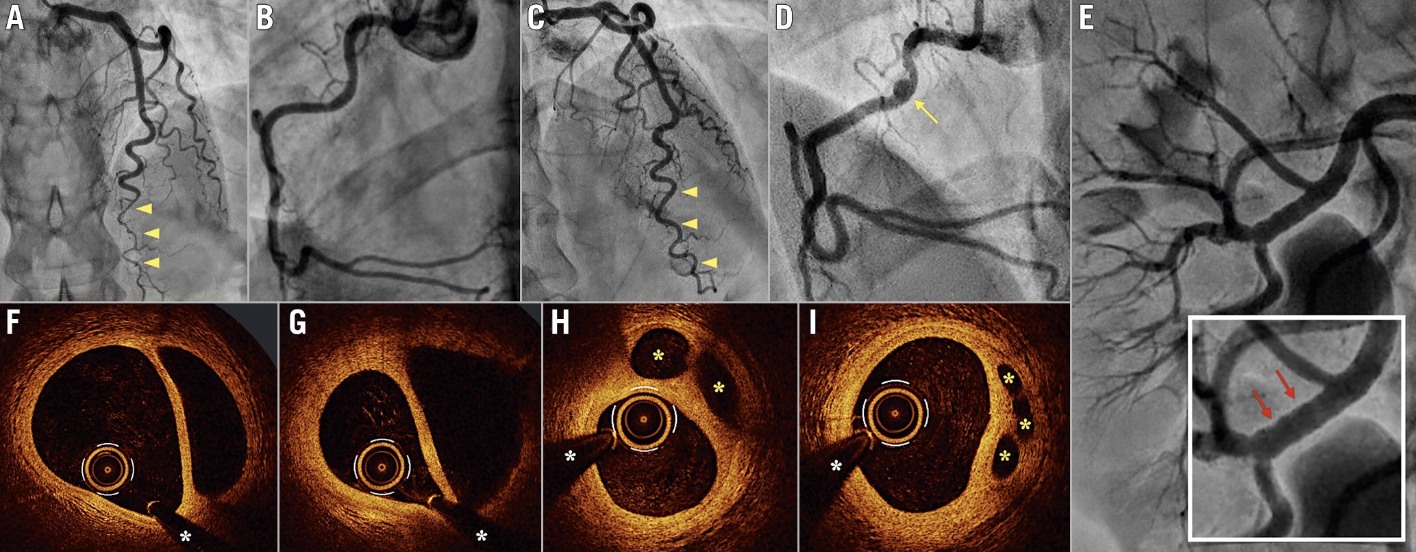
Figure 1. Coronary angiographic and OCT images. A) Coronary angiography showing a diffuse lumen narrowing of the distal LAD artery (yellow arrowheads) with no stenosis in the RCA (B). C) Coronary angiography showing complete healing of the distal LAD (yellow arrowheads) and the proximal RCA with mild stenosis and a small aneurysm (D, yellow arrow). E) Angiography displaying stigmas of fibromuscular dysplasia in the right renal artery (red arrows). F,G) Optical coherence tomography images depicting the characteristic double-lumen pattern, and (H,I) the “lotus root” appearance, with multiple thick intimal septa splitting the false lumen into small, rounded channels in the proximal RCA (yellow asterisks). White asterisk denotes wire artefact. LAD: left anterior descending coronary artery; RCA: right coronary artery
Conflict of interest statement
The authors have no conflicts of interest to declare concerning this study.
Supplementary data
To read the full content of this article, please download the PDF.
Moving image 1. Coronary angiography of the left anterior descending artery (first procedure).
Moving image 2. Coronary angiography of the right coronary artery (first procedure).
Moving image 3. Coronary angiography of the left anterior descending artery (second procedure).
Moving image 4. Coronary angiography of the right coronary artery (second procedure).
Moving image 5. OCT cross-sectional pullback showing the “lotus root” appearance in the proximal right coronary artery.