IMAGE IN CARDIOLOGY

DOI: 10.4244/EIJ-D-16-00614

Single coronary artery arising from the right aortic sinus with dual LAD supply from separate branches

Christopher V. DeSimone, MD, PhD; Eric Williamson, MD; Hartzell V. Schaff, MD; Gregory W. Barsness, MD; Scott J. Hoffman, MD; Verghese Mathew*, MD

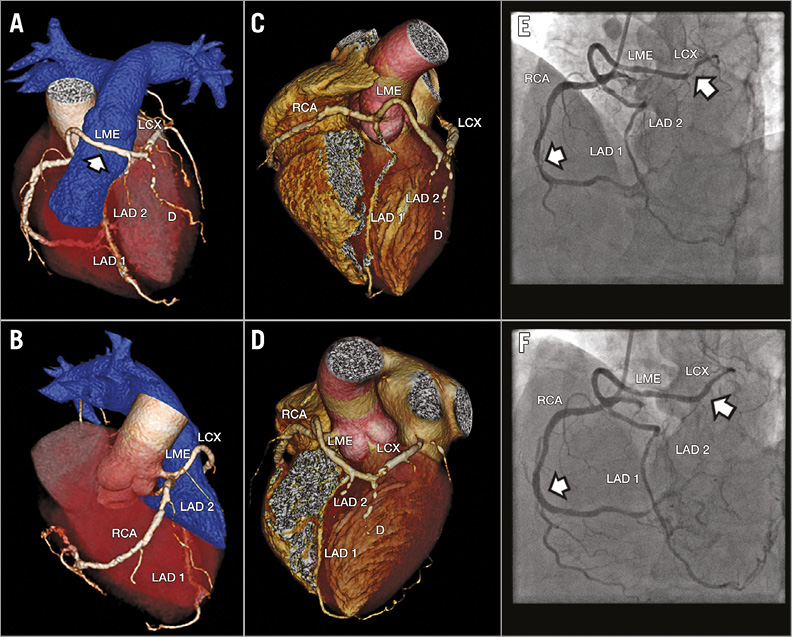
A 55-year-old male presented with angina and reversible ischaemia on stress imaging. He had a rare combination of an anomalous single coronary artery, dual left anterior descending artery (LAD) supply from 1) a transseptal, long middle/distal LAD, and 2) a separate, pre-pulmonic, short left main equivalent supplying the proximal LAD, septal, diagonal, and left circumflex (LCX) territories. CT reconstruction (Panels A-D) demonstrated a single coronary artery from the right aortic sinus which trifurcated into the right coronary system and two additional branches supplying the left coronary system. One branch travelled along the right ventricular (RV) aspect of the septum as a long middle and distal LAD equivalent and supplied branches along the inferolateral and lateral left ventricle. A second branch served as a left main equivalent with a pre-pulmonic course anterior to the RV outflow tract, prior to giving rise to a small proximal LAD and associated septal and diagonal branches, and LCX (Panel A: LAO cranial view; Panel B: LAO caudal view; Panel C: RAO cranial view; Panel D: LAO cranial view. RCA: right coronary artery; LME: left main equivalent; LCX: left circumflex artery. LAD 1: LAD coursing intraseptally; LAD 2: arising from LME; D: diagonals. White arrow: LME coursing anterior to the pulmonary trunk). The patient underwent successful PCI of the mid-RCA and proximal LAD (Panels E & F: LAO cranial view; arrowheads pointing to stenotic lesions pre- and post-stent).

Conflict of interest statement

The authors have no conflicts of interest to declare.
Volume 12 Number 17
Apr 20, 2017
Volume 12 Number 17
View full issue


Key metrics

Suggested by Cory

IMAGE IN CARDIOLOGY

10.4244/EIJ-D-16-00393 Apr 20, 2017
Multiple spontaneous intimal dissections with single left coronary artery and coronary artery aneurysm
Achkouty G et al
free

IMAGE IN CARDIOLOGY

10.4244/EIJV11I13A291 Apr 20, 2016
A disaster never comes alone: total ostial occlusion of the left main coronary artery with an anomalous origin
Rodrigues P et al
free

10.4244/EIJV9I11A228 Mar 20, 2014
How should I treat a coronary bifurcation wide-neck aneurysm?
Sgueglia G et al
free
Trending articles
151.43

State-of-the-Art

10.4244/EIJ-D-22-00776 Apr 3, 2023
Computed tomographic angiography in coronary artery disease
Serruys PW et al
free
55.9

Clinical research

10.4244/EIJ-D-22-00621 Feb 20, 2023
Long-term changes in coronary physiology after aortic valve replacement
Sabbah M et al
free
54.9

Expert review

10.4244/EIJ-D-21-01010 Jun 24, 2022
Device-related thrombus following left atrial appendage occlusion
Simard T et al
free
43.75

Clinical Research

10.4244/EIJ-D-21-01091 Aug 5, 2022
Lifetime management of patients with symptomatic severe aortic stenosis: a computed tomography simulation study
Medranda G et al
free
39.95

Clinical research

10.4244/EIJ-D-22-00558 Feb 6, 2023
Permanent pacemaker implantation and left bundle branch block with self-expanding valves – a SCOPE 2 subanalysis
Pellegrini C et al
free
X

The Official Journal of EuroPCR and the European Association of Percutaneous Cardiovascular Interventions (EAPCI)

EuroPCR EAPCI
PCR ESC
Impact factor: 7.6
2023 Journal Citation Reports®
Science Edition (Clarivate Analytics, 2024)
Online ISSN 1969-6213 - Print ISSN 1774-024X
© 2005-2024 Europa Group - All rights reserved