Coronary agenesis is defined as the absence of either the right or the left coronary ostium. We present the case of a 40-year-old man who underwent elective percutaneous coronary intervention (PCI) of a right coronary ostium agenesis (Figure 1A). Baseline coronary angiography showed a normal left coronary artery (LCA) and right coronary artery (RCA) ostium agenesis receiving blood flow from the LCA (Figure 1B, Moving image 1). A 0.014 inch Pilot 50 (Abbott Vascular, Santa Clara, CA, USA) guidewire successfully passed through a very tortuous conus branch with a 150 cm FineCross® (Terumo Corp., Tokyo, Japan) microcatheter support (Figure 1C). A 0.014 inch ULTIMATEbros3 (ASAHI Intecc, Aichi, Japan) guidewire successfully punctured through the RCA agenesis and floated inside the ascending aorta (Moving image 2). Snaring of the guidewire was done using an EN Snare® (Merit Medical, Galway, Ireland). Volcano intravascular ultrasound (IVUS) (Kimball, Tampa, FL, USA) revealed the ostium of the RCA without visible coronary structure (Figure 1D). A biolimus-eluting stent (3.0×24 mm) was deployed at the ostium of the RCA (Biosensors Europe SA, Morges, Switzerland). Final angiography revealed a good result (Moving image 3). This case demonstrates retrograde approach PCI for a congenital RCA ostium agenesis. IVUS documented the true vessel anatomy and the agenesis vessel anatomy. Accordingly, utilisation of the CTO technique allowed revascularisation of the coronary artery agenesis without complication.
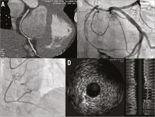
Figure 1. The images of coronary agenesis. A) Multislice cardiac computed tomography showed an RCA ostium agenesis. B) Coronary angiogram showed an RCA ostium agenesis receiving blood flow from LCA. C) The 0.014 inch Pilot 50 passed the tortuous conus branch with a 150 cm FineCross microcatheter. D) IVUS showed the structure of the proximal RCA vessel anatomy with ostium agenesis.
Conflict of interest statement
The authors have no conflicts of interest to declare.
Online data supplement
Moving image 1. Baseline coronary angiogram.
Moving image 2. Retrograde ULTIMATEbros3 guidewire floating inside ascending aorta.
Moving image 3. Final coronary angiogram.
Supplementary data
To read the full content of this article, please download the PDF.
Moving image 1. Baseline coronary angiogram.
Moving image 2. Retrograde ULTIMATEbros3 guidewire floating inside ascending aorta.
Moving image 3. Final coronary angiogram.