The success of transcatheter aortic valve implantation (TAVI) has had a huge impact on the management of patients with aortic stenosis1. At the heart of it is a basic idea of implanting a heart valve which does not need sutures and is held securely by tissue because of relative oversizing. Some of the issues associated with the first two transcatheter heart valve (THV) devices, i.e., SAPIEN (Edwards Lifesciences, Irvine, CA, USA), and CoreValve® (Medtronic, Minneapolis, MN, USA) were the inability to reposition, paravalvular leaks and conduction disturbances1. Second- and third-generation THV designs were introduced to address some of these issues and thus novel concepts and designs to allow repositioning and recapturability were introduced. The Direct Flow Medical® (DFM) valve (Direct Flow Medical, Santa Rosa, CA, USA) and the Lotus™ (Boston Scientific, Marlborough, MA, USA) embody such concepts, allowing a retrograde delivery of the device, essentially through the femoral artery into the aortic position2.
In parallel, the use of a THV to treat a degenerated surgical heart valve, i.e., valve-in-valve (VIV), has been a natural extension of TAVI3. Today there is a substantial body of experience in treating degenerated surgical valves, predominantly in the aortic position but also in mitral, tricuspid and pulmonary positions. Similarly, reports have now emerged of treating patients with failing mitral valve repair with THV prostheses. Mitral valve repair is an excellent treatment but the reported failure rate is in the range of 10% at five years and up to 30% at 10 years4. An annuloplasty ring is employed in the majority of mitral repairs and is used to provide an anchor for the THV4. Kempfert and colleagues first demonstrated the feasibility, in an animal model, of the use of the SAPIEN valve in a semi-rigid ring5. Since then, there have been multiple reports of successful valve-in-ring (VIR) procedures, all of them using the SAPIEN valve, which is a balloon-expandable valve and hence not repositionable or retrievable6,7. To date, the largest single-centre experience was recently reported by Bouleti and colleagues7. They treated 11 patients (nine Edwards Physio and two Medtronic Duran AnCore® rings). Ring sizes ranged from 26 to 36 mm and SAPIEN sizes 23, 26 and 29 mm were used, depending on the ring size and CT dimensions. A successful result was obtained in nine patients; two patients had moderate residual paraprosthetic regurgitation and had to be operated within six months. Three patients developed left ventricular outflow tract obstruction (LVOTO) but were treated conservatively.
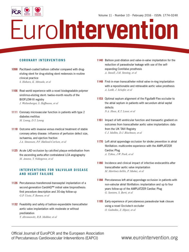
In this issue, Latib and co-authors describe the first-in-man experience of the use of a repositionable and retrievable DFM valve to treat failed mitral repairs8. The total experience reported is of eight cases in four centres. All cases were carried out under general anaesthesia through a transapical approach. All cases were discussed by the Heart Team and were thought to be high risk for reoperative surgery (average logistic EuroSCORE was 36.5±17.9). All patients had semi-rigid rings (two Edwards Physio, two SJM™ Séguin [St. Jude Medical, St. Paul, MN, USA] and four Medtronic CG Future® Ring). The sizes of the rings treated were 26, 28, 30 and 34 mm. The DFM valves used were 25, 27 and 29 mm. The DFM valve size chosen was at least 25% more than the mitral ring area, as indicated by the manufacturer. The valve was positioned and assessment carried out for the presence of paravalvular leak, adequate fit of the valve and LVOTO. In two cases the DFM valve was not implanted as a significant paravalvular leak became evident in one, and in the other there was significant LVOTO. The first patient underwent open surgery successfully and the second patient an alcohol septal ablation. Of the six implants, one patient had significant LVOTO, which was treated by repositioning the device more atrially. In another patient, a size 25 DFM valve was implanted but was loose and hence exchanged for a 27 DFM valve with a good result. Overall, the results were satisfactory and in 50% of patients they used the ability either to reposition or to retrieve the device to achieve a good end result.
There are four aspects which need to be considered before undertaking a VIR procedure and which determine the outcome:
1. characteristics of the ring,
2. characteristics of the THV including the delivery system,
3. probability of LVOTO, and
4. possibility of delayed embolisation.
There are multiple types of ring used for mitral repair9. These may be classified as bands and rings. Bands can be incomplete or complete depending on whether they are sutured only to the posterior annulus or to the posterior and anterior annulus, respectively. Bands may not provide a secure anchor for THV implantation. This is an important consideration before undertaking a VIR procedure. Mitral rings tend to be complete, except for a few where there is a small gap anteriorly, but more importantly they can be either rigid or semi-rigid (semi-flexible). This property determines whether the ring can be forced into a circular or near circular shape after THV implantation. By and large, rigid rings retain their original non-circular shape and hence tend to deform the THV, while semi-rigid rings tend to become circular or near circular and hence they are more suited for a VIR procedure as they result in less deformation of the THV. One of the other important considerations is the size of the mitral ring. Rings greater than size 32 tend to be too large for current THV devices, as a degree of oversizing is essential for secure THV placement6-9.
The design of the THV and its delivery system also influence its use for mitral VIR procedures. For example, only shorter THV devices can be implanted in the mitral position, e.g., SAPIEN, Lotus and DFM valves2. While the SAPIEN can be implanted either in the antegrade or retrograde manner, the DFM valve and the Lotus can only be implanted in a retrograde manner (transapical route) as they are attached to their respective delivery systems. It should be kept in mind that the ability of a THV to deform the ring to a circular shape varies from design to design. The balloon-expandable SAPIEN valve has the maximum ability to force the ring into a circular shape but when used in rigid rings will result in valve deformation, while the DFM and Lotus may not have as much ability to circularise the ring but can adapt and function better in a non-circular ring6-9. It is too early to say whether the ability to force the ring into a circular shape or to adapt to the non-circular shape of the ring is the better strategy for long-term durability. The largest experience in VIR has been with the balloon-expandable valve (SAPIEN, SAPIEN XT and SAPIEN 3). Although initial experience especially in treating semi-rigid rings has been satisfactory, reports of early degeneration and residual PV leaks have started to emerge7,10. One of the other disadvantages of using a balloon-expandable valve is the inability to reposition and recapture. This could be important if the device was deemed to be too big or small or if, after implantation, a significant LVOTO was observed7. Repositionable and recapturable devices provide the operator with a chance to “test drive” and correct before the device is released. This feature was useful in two out of eight cases reported by Latib and colleagues where, in the first case, a significant paravalvular leak became obvious and hence the device was removed and open surgery performed, and in the second patient LVOTO was observed and hence the device was repositioned more atrially before the final implant8.
The possibility of LVOTO is real after a VIR procedure11. This is because the anterior mitral leaflet is hinged open (similar to a fixed systolic anterior motion) because of the THV7,8. The aetiology of LVOTO is multifactorial, but device-related factors are more ventricular deployment and larger ventricular flare of the device (directly proportional to oversizing)11. The other problem with mitral VIV (also theoretically possible with VIR) is delayed embolisation of the THV device12. To prevent this, one must have a degree of oversizing, but on the other hand too much oversizing may lead to LVOTO. Hence, this is a delicate balance and each procedure should be individualised. This is partly obvious from the reported series, as two patients who developed LVOTO had 80% oversizing of the DFM prosthesis: one responded to repositioning but in the other the device had to be removed. What is interesting and evident from the report is that in two other patients the oversizing was >80% but they did not develop LVOTO. This could be because of favourable factors related to the left ventricle such as cavity size, septal bulge and aortomitral annular angle11.
To summarise, the VIR procedure using existing THV devices seems feasible but involves unique challenges and limitations. Partly this is to do with the ring design, which may not be compatible with a THV device, but is also due to factors still not well understood, i.e., LVOTO and delayed embolisation. We also need to switch our focus from the implantability of devices to their durability. While one can anticipate good results if the ring is suitable, the THV device is deployed in a near circular shape and there is no LVOTO; one can anticipate a bad result (early or intermediate failures) if this is not achieved, or the result can be ugly if there is complete deformation of the THV device, significant LVOTO or embolisation resulting in intraprocedural or early haemodynamic compromise. Hence, VIR should only be considered in patients when surgery is considered high risk, when the ring is suitable for a VIR procedure and when there is confidence in the positive midterm outcome.
Conflict of interest statement
V. Bapat is consultant for Edwards Lifesciences, Boston Scientific and Sorin.

