Introduction
Transcatheter aortic valve implantation (TAVI) is now an established alternative treatment for patients with severe calcific aortic stenosis deemed at high risk for conventional surgical aortic valve replacement1. Tens of thousands of procedures have been performed worldwide since the first implant by Cribier in 2002. As the number of procedures continues to increase rapidly, the procedures and devices have evolved, leading to improvements in patient outcomes. Enthusiasm for the technology has translated into increased use in the treatment of failing bioprosthetic valves. This use was first reported by Wenaweser and colleagues in 20072 and offers an alternative to high-risk redo valve surgery. Although the first procedure was reported in a failing prosthesis in the aortic position, similar procedures have been reported in mitral, pulmonary and tricuspid positions. The valve-in-valve (VIV) procedure does, however, come with a distinct set of technical challenges and considerations including correct valve sizing, positioning, predicting and avoiding coronary obstruction, the challenge of prosthesis-patient mismatch, and minimising paravalvular regurgitation and permanent pacing. Establishing long-term durability of the device also remains a challenge.
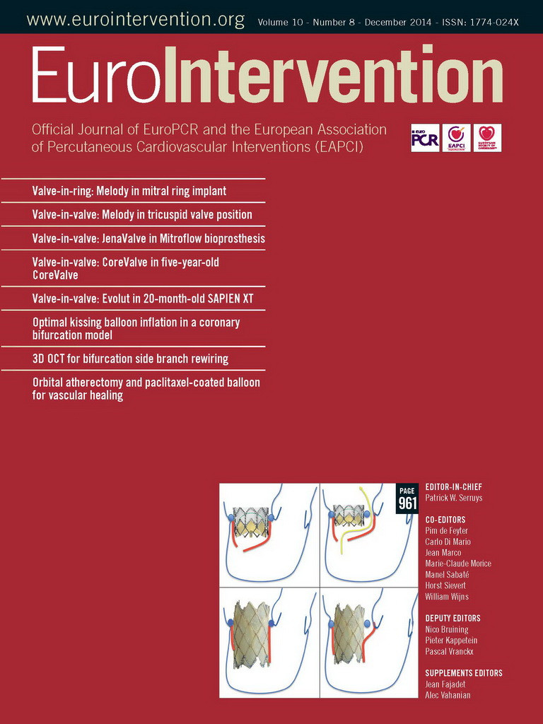
In this issue of EuroIntervention, four contributions explore the VIV procedure. Firstly, Tzifa et al have reported on their series of VIV implantation in failing prostheses in the tricuspid position3. Conradi et al have addressed the challenge of treating the Mitroflow bioprosthesis (Sorin, Milan, Italy)4. Lastly, we have two images, one from Beatriz Vaquerizo beautifully demonstrating the safety and five-year durability of failing transcatheter aortic valve-in-transcatheter aortic valve implantation5, and the second from Ran Kornowski showing the medium-term results of a failing balloon-expandable valve treated with a self-expandable valve6.
Tricuspid valve-in-valve
Primary tricuspid valve disease is rare, and therefore tricuspid valve replacement is not a common operation. The right side of the heart provides a unique set of anatomical and functional considerations and, unfortunately, tricuspid valve replacement is still associated with high postoperative mortality7. Consequently, experience in managing failing prostheses in this position is limited. However, the risk involved in redo operations in this cohort of patients is often prohibitively high, and therefore transcatheter approaches are likely to evolve as an attractive treatment of choice. The current report of five patients treated by a VIV implantation by Tzifa and colleagues represents the second largest experience published3. The bioprosthetic valves were severely regurgitant in four patients, two of whom also had moderate-severe stenosis, while the other patient had severe stenosis. Transcatheter treatment was with the Melody® valve (Medtronic, Minneapolis, MN, USA) in four patients, and the Edwards SAPIEN valve (Edwards Lifesciences, Irvine, CA, USA) in one. The right internal jugular was the favoured approach in the majority of cases with one being deployed transfemorally. Correct sizing of the device was ensured by balloon sizing as well as transoesophageal echocardiography in all patients –important in avoiding oversizing of the prosthesis, which can affect the conduction tissue which is in close proximity to the valve, as well as valvular regurgitation. There were no complications recorded and all patients were discharged within 24 hours. During a follow-up of 15-22 months, all patients had significant improvement in valvular function and peripheral oedema, as well as NHYA class.
The authors have to be congratulated on this important report. It does however highlight that these are rare procedures. The cumulative experience of five implants was from three very high-volume congenital and adult structural intervention teams. The largest report of tricuspid VIV in 15 patients was pooled from eight centres8. There is also a string of case reports which have been published on the procedure, but reporting bias may prevent us from appreciating fully the technical challenge of achieving a good outcome in this cohort of patients. However, the principles of correct valve sizing and positioning are common fundamental goals in VIV procedures, and experience in other valve positions should translate into improved outcomes.
Aortic valve-in-valve
Since the first report of an aortic VIV procedure by Wenaweser in 20072, several hundred procedures have been performed worldwide. The most comprehensive assessment of the procedure was published by Danny Dvir this year, reporting on a registry of 459 such procedures using the self-expandable CoreValve (Medtronic) and balloon-expandable Edwards SAPIEN devices (Edwards Lifesciences)9. The major mechanism of failure of the existing prosthesis was evenly distributed among regurgitation, stenosis and combined. Factors associated with worse outcomes were the presence of surgical valve stenosis and small bioprostheses/prosthesis-patient mismatch9. Correct valve sizing is of critical importance. Most operators have used the stent internal diameter of the surgical heart valve to select the appropriate transcatheter valve size. Unfortunately, the true internal diameter in valves sutured inside the stent is often smaller, by approximately 2 mm for porcine valves, and 1 mm for pericardial valves10. This is especially important in borderline sizes in order to avoid the problems associated with oversizing, as well as confirming the suitability of the procedure in the smaller label sizes. Correct positioning of the valve is also extremely important in optimising outcomes, specifically for the native prosthesis as well as the proposed implant device. A smartphone application addressing each aspect of the VIV procedure, including the specifics of the existing prosthesis such as design and suitability for VIV treatment, sizing (including height and true internal diameter), and suggested prosthesis and optimal positioning, has been developed for aortic VIV as well as mitral valve-in-ring, which provides valuable information even in experienced hands11. The improvement of our understanding of each of these aspects of the procedure has already made the VIV procedure very predictable, with favourable outcomes. This has also impacted on surgical practice where there is a developing trend towards implanting bioprostheses in younger patients12, with the less invasive treatment option of VIV for potential prosthesis failure in the future.
Mitroflow bioprosthesis
An ongoing challenge in VIV procedures is the potential for coronary obstruction, which is a major predictor of adverse outcome. With improvement in the number and size of available prostheses, and understanding of the optimal positioning of the implantation, this situation has improved. However, certain surgical prostheses have a greater tendency for coronary obstruction during VIV. One of these is the Mitroflow (Sorin), which has an over eight times greater incidence of ostial coronary obstruction compared to other stented bioprostheses13. This is predominantly due to the fact that the leaflets are mounted on the outside of the valve stent, as well as a relatively tall valve in height. What makes this issue of particular importance is the prevalence of this implant in surgical practice, and therefore for VIV procedures: it accounted for approximately 20% of the valves treated with VIV in the global registry13. Further, smaller sizes of Mitroflow are implanted in small aortic roots, which is an unfavourable anatomy for a VIV procedure. The Mitroflow has been treated with balloon-expandable devices such as the Edwards SAPIEN, and self-expandable devices such as the Medtronic CoreValve and St. Jude Portico (St. Jude Medical, St. Paul, MN, USA). In this issue of EuroIntervention, Conradi and colleagues report on two cases of failing Mitroflow bioprostheses, both due to severe regurgitation, using the JenaValve (JenaValve Technology GmbH, Munich, Germany)4. The surgical valves were both 25 mm, with true internal diameters of 21 mm, which were treated with 23 mm JenaValves. The use of the JenaValve in this situation provides a new paradigm, where the native leaflets are clipped to the valve stent, thereby potentially eliminating the risk of coronary occlusion. Both cases resulted in successful implantation of the VIV prosthesis with improvement of clinical symptoms and no complications noted. However, both cases had significant residual gradients of 55/22 and 38/22 (peak/mean mmHg), likely to have arisen from the unrestricted outer diameter of the JenaValve which is in the region of 28 mm with the true internal diameter of the Mitroflow being 21 mm. The impact of significant residual valve stenosis on the durability of the procedure is likely to be an important factor but remains to be established. Another option for such situations would be to use a smaller THV to avoid excessive oversizing, which in turn leads to outward deflection of stent posts and leaflets, increasing chances of coronary obstruction. Until recently the 23 mm THV was the smallest valve available but now 20 mm SAPIEN XT (commercial) and 20 mm SAPIEN 3 (compassionate use only) are available and will be ideal for valves with a true ID less than 19 mm. This could also result in a more circular deployment of the SAPIEN valve and hence better function.
Durability of valve-in-valve
A remaining issue of critical importance is the durability of the VIV procedure, particularly due to the fact that, on average, patients undergoing VIV are younger and have fewer comorbidities than high-risk patients who have TAVI as their index procedure. Data addressing this issue will slowly emerge as longer follow-up data become available. This will allow us to identify further patient/procedural/prosthetic factors that are associated with adverse outcome, which should aid in further refinement of these procedures in order to continue to optimise patient outcomes.
Conclusion
The valve-in-valve procedure has provided an important gateway to avoiding high-risk redo surgery and is now a potential option for all surgically implanted valves or rings in all valve positions. The number of these procedures is likely to expand rapidly, as younger and younger patients are implanted with bioprosthetic valves, and the exponential growth of TAVI results in the likely increasing burden of failing transcatheter valves. Adequate planning, prosthesis selection and correct placement of the device continue to be central to a successful implantation.
Conflict of interest statement
V. Bapat is a consultant for Edwards Lifesciences, Medtronic, and St. Jude Medical. K. Asrress has no conflicts of interest to declare.

