DOI: 10.4244/EIJV8I3A62

Left ventricular angiography with a Jacky Radial catheter at high pressure leading to myocardial staining and opacification of the great cardiac vein and coronary sinus

Gurjaipal Kang1*, MD, UPMC; Kevin Kang1; Javier Jurado2, MD; Luke Byrnes2, MD

A 66-year-old female presented with chest discomfort, diagnosed as Canadian class III stable angina. Her Lexiscan nuclear stress test showed normal perfusion but a reduced ejection fraction of 45%. A coronary angiogram revealed minimal luminal disease except for a diagonal lesion. A left ventricular angiogram using 30 ml of contrast over two seconds with a Jacky Radial catheter with side holes at 600 pound square inch revealed an anterior myocardial stain. There was immediate antegrade filling of the anterior inter-ventricular vein adjoining the myocardial stain. Both the great cardiac vein and the coronary sinus emptying into the right atrium were also seen. The American Board certified interventional cardiologist at the time thought that this was the left anterior descending artery and unnecessarily again injected the left main selectively to document normal flow and lack of perforation. Diagnostic difficulty was therefore noted due to this unusual staining and dense coronary venous opacification. Caution should be exercised during left ventricular angiography so that myocardial injection is avoided because it causes arrhythmias, pain, rupture and death. The end-hole of a catheter should not be abutting the myocardium, and in those situations with a catheter having a pointing end-hole, low pressures for injection should be used to avoid myocardial injury.
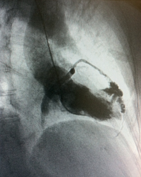
Figure 1. Coronary venous filling and myocardial stain.

Conflict of interest statement

The authors have no conflicts of interest to declare.

Volume 8 Number 3
Jul 27, 2012
Volume 8 Number 3
View full issue


Key metrics

Suggested by Cory

IMAGE IN CARDIOLOGY

10.4244/EIJV11I7A169 Nov 20, 2015
An atypical clinical presentation of a broken guidewire left in the venous system
Montero-Cabezas J et al
free

INTERVENTIONAL FLASHLIGHT

10.4244/EIJ-D-17-00048 Jun 20, 2017
Iatrogenic coronary artery dissection induced during invasive absolute coronary blood flow measurement: optical coherence tomography findings
García-Guimaraes M et al
free

10.4244/EIJV8I1A23 May 15, 2012
How should I treat guidewire-induced distal coronary perforation?
Senguttuvan N et al
free

IMAGE IN CARDIOLOGY

10.4244/EIJV11I13A291 Apr 20, 2016
A disaster never comes alone: total ostial occlusion of the left main coronary artery with an anomalous origin
Rodrigues P et al
free
Trending articles
200.45

State-of-the-Art

10.4244/EIJ-D-21-00089 Jun 11, 2021
Intracoronary optical coherence tomography: state of the art and future directions
Ali ZA et al
free
154.43

State-of-the-Art

10.4244/EIJ-D-22-00776 Apr 3, 2023
Computed tomographic angiography in coronary artery disease
Serruys PW et al
free
92.95

State-of-the-Art Review

10.4244/EIJ-D-20-01296 Aug 27, 2021
Management of cardiogenic shock
Thiele H et al
free
47.4

State-of-the-Art

10.4244/EIJ-D-24-00386 Feb 3, 2025
Mechanical circulatory support for complex, high-risk percutaneous coronary intervention
Ferro E et al
free
43.65

Clinical research

10.4244/EIJ-D-23-00590 Dec 4, 2023
Prognostic impact of cardiac damage staging classification in each aortic stenosis subtype undergoing TAVI
Nakase M et al
free
36.5

State-of-the-Art

10.4244/EIJ-D-23-00448 Jan 15, 2024
Coronary spasm and vasomotor dysfunction as a cause of MINOCA
Yaker ZS et al
free
34.75

State-of-the-Art

10.4244/EIJ-D-23-00606 Jan 1, 2024
Targeting inflammation in atherosclerosis: overview, strategy and directions
Waksman R et al
free
X

The Official Journal of EuroPCR and the European Association of Percutaneous Cardiovascular Interventions (EAPCI)

EuroPCR EAPCI
PCR ESC
Impact factor: 7.6
2023 Journal Citation Reports®
Science Edition (Clarivate Analytics, 2024)
Online ISSN 1969-6213 - Print ISSN 1774-024X
© 2005-2025 Europa Group - All rights reserved