IMAGE IN CARDIOLOGY

DOI: 10.4244/EIJV11I7A169

An atypical clinical presentation of a broken guidewire left in the venous system

Jose M. Montero-Cabezas*, MD; Robin de Groot, MD; Martin J. Schalij, MD, PhD

A 65-year-old male patient with a non-ischaemic cardiomyopathy with poor left ventricular function was referred for right-left heart catheterisation as part of the work-up for left ventricular assist device implantation.

Using a femoral approach, a Swan-Ganz (SG) catheter (Edwards Lifesciences, Irvine, CA, USA) was advanced into the right atrium and, subsequently, to the right ventricle. After several unsuccessful attempts at advancing it into the pulmonary artery, a 0.014” 300 cm BHW coronary guidewire (Abbott Vascular, Santa Clara, CA, USA) was advanced through the catheter, allowing it to be advanced into the pulmonary artery. After pressure recording, the catheter was removed and the procedure finished without complications.

A few hours later, the patient referred to a mild throbbing discomfort localised at the fifth left intercostal space at the level of the midclavicular line. Physical examination revealed a pulsatile point in which a sharp object seemed to be protruding under the skin (Figure 1A, Moving image 1). Fluoroscopic examination showed a long guidewire fragment stabbing the apex of the right ventricle (Figure 1B, Moving image 2). The wire was removed by using an Amplatz GooseNeck® snare (ev3 Inc., Plymouth, MN, USA) via the right femoral vein (Figure 1C).

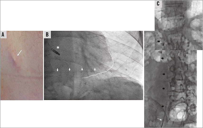
Figure 1. Broken guidewire identification and retrieval. A) Pulsatile point. B) Dotted line: cardiac silhouette. White arrows: guidewire fragment fixed to the apex of the right ventricle. Asterisk: MitraClip® device (Abbott Vascular). C) Retrieval of the guidewire fragment (white arrow). Dotted arrow: Amplatz GooseNeck® snare. Black arrows: snare catheter.

This extraordinary clinical presentation led to a prompt identification of the complication and its successful resolution.

Conflict of interest statement

The authors have no conflicts of interest to declare.

Supplementary data

Moving image 1. Pulsatile point.

Moving image 2. Guidewire fragment embedded in the right ventricle.

Supplementary data

To read the full content of this article, please download the PDF.

Pulsatile point.

Moving image 2. Guidewire fragment embedded in the right ventricle.

Volume 11 Number 7
Nov 20, 2015
Volume 11 Number 7
View full issue


Key metrics

Suggested by Cory

IMAGE IN CARDIOLOGY

10.4244/EIJY14M07_07 Jul 20, 2015
Percutaneous extraction of pacing leads from the left coronary artery and left ventricle
Elmariah S et al
free

10.4244/EIJV10I61A131 Oct 20, 2014
How should I treat an accidentally misplaced 8 Fr drainage catheter in the right ventricle?
Bakos Z and Harnek J
free

Jan 15, 2010
Femoral artery puncture
Haude M et al
free
Trending articles
318.1

State-of-the-Art Review

10.4244/EIJ-D-21-00695 Nov 19, 2021
Transcatheter treatment for tricuspid valve disease
Praz F et al
free
117

State-of-the-Art

10.4244/EIJ-D-24-00066 Apr 21, 2025
Management of complications after valvular interventions
Bansal A et al
free
108.3

Viewpoint

10.4244/EIJ-E-22-00007 May 15, 2022
TAVI at 20: how a crazy idea led to a clinical revolution
Eltchaninoff H et al
free
91.6

Image – Interventional flashlight

10.4244/EIJ-D-22-00344 Aug 5, 2022
First dedicated transcatheter leaflet splitting device: the ShortCut device
Tchétché D et al
free
71.3

State-of-the-art

10.4244/EIJ-D-22-00627 Feb 6, 2023
Left atrial appendage occlusion
Holmes D et al
free
68.9

State-of-the-Art

10.4244/EIJ-D-24-00992 Sep 15, 2025
Antithrombotic therapy in complex percutaneous coronary intervention
Castiello D et al
free
60.65

Clinical research

10.4244/EIJ-D-20-01155 Oct 20, 2021
A deep learning algorithm for detecting acute myocardial infarction
Liu W et al
free
56.7

Expert Review

10.4244/EIJ-D-25-00201 Oct 10, 2025
Drug-coated balloons for coronary bifurcation lesions
Fezzi S et al
free
56.7

Expert Review

10.4244/EIJ-D-25-00201 Oct 20, 2025
Drug-coated balloons for coronary bifurcation lesions
Fezzi S et al
free
49.55

CLINICAL RESEARCH

10.4244/EIJ-D-17-00962 Apr 6, 2018
A new optical coherence tomography-based calcium scoring system to predict stent underexpansion
Fujino A et al
free
X

PCR
Impact factor: 9.5
2024 Journal Citation Reports®
Science Edition (Clarivate Analytics, 2025)
Online ISSN 1969-6213 - Print ISSN 1774-024X
© 2005-2025 Europa Group - All rights reserved