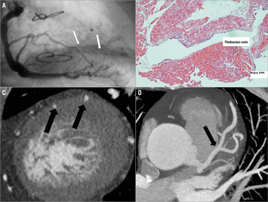
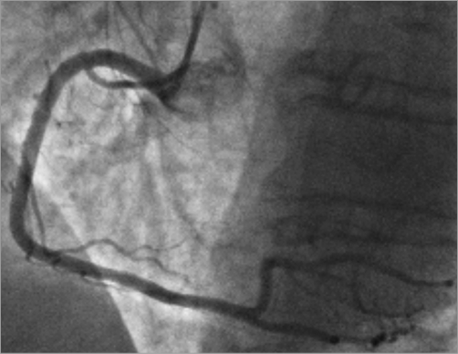
We describe two cases of direct coronary drainage to the left ventricle via Thebesian veins detected during coronary angiography with contrast progressing into the myocardium via the microcirculation ending in the left ventricular cavity. First, an 86-year-old male cardiac transplant patient in whom the right coronary artery showed left ventricular opacification via Thebesian veins (Panel A, Moving image 1, Moving image 2, Online Figure 1). Unlike the biopsy from the right ventricle (Online Figure 2), the left ventricular biopsy shows the presence of Thebesian veins (Panel B). Second, a 77-year-old female with chest pain (Moving image 3, Moving image 4, Online Figure 3, Online Figure 4). The cardiac CT shows the intramyocardial course of coronary arteries (Panel C), coronary enlargement (Panel D) and a heterogeneous contrast distribution within the left and right ventricular myocardium (Online Figure 5).
Thebesian veins form the smaller and often underappreciated cardiac vein circulation that drain the blood directly into the ventricular cavities. Exclusively left ventricular drainage through Thebesian veins is a rare and impressive congenital abnormality without clinical implications.
Conflict of interest statement
J. Adjedj declares having received a research grant from the Fédération Francaise de Cardiologie. The other authors have no conflicts of interest to declare.
Supplementary data
Moving image 1. 86-year-old male patient. The left coronary artery shows direct coronary drainage into the anterolateral part of the left ventricle.
Moving image 2. The same patient. The right coronary artery shows direct coronary drainage into the inferior part of the left ventricle.
Moving image 3. 77-year-old female patient. The left coronary artery shows direct coronary drainage into the whole left ventricle.
Moving image 4. The same patient. The right coronary artery has a normal drainage of contrast.
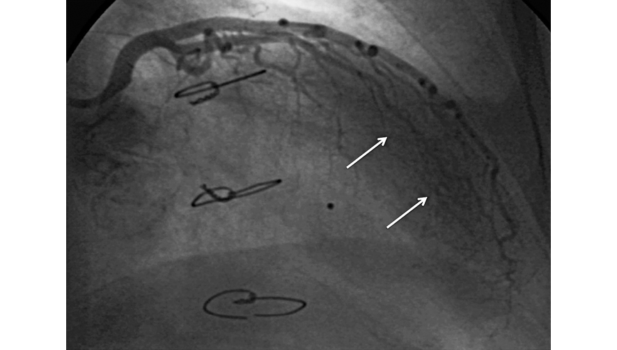
Online Figure 1. Left coronary artery with opacification of the LV via Thebesian veins (arrows).
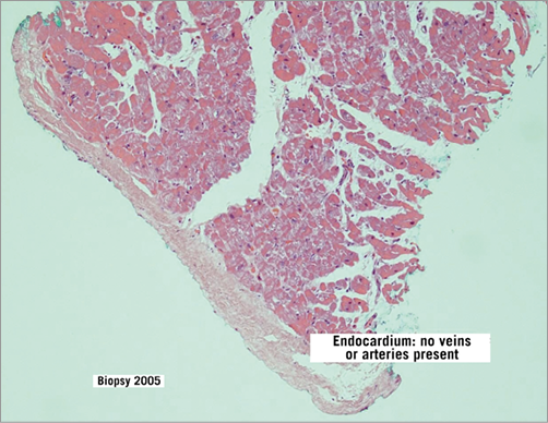
Online Figure 2. Right ventricular biopsy without presence of Thebesian veins.
Online Figure 3. RCA without opacification of the LV via Thebesian veins.
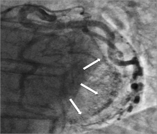
Online Figure 4. LCA with opacification of the LV via Thebesian veins (arrows).
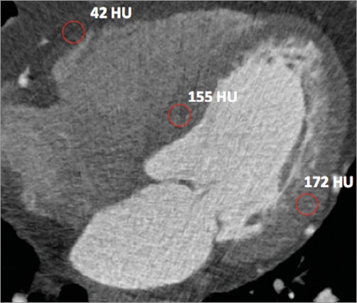
Online Figure 5. Myocardial brightness differences between left, septal and right myocardium (circles). The brightness level is expressed in Hounsfield units (HU).
Supplementary data
To read the full content of this article, please download the PDF.
Moving image 1. 86-year-old male patient. The left coronary artery shows direct coronary drainage into the anterolateral part of the left ventricle.
Moving image 2. The same patient. The right coronary artery shows direct coronary drainage into the inferior part of the left ventricle.
Moving image 3. 77-year-old female patient. The left coronary artery shows direct coronary drainage into the whole left ventricle.
Moving image 4. The same patient. The right coronary artery has a normal drainage of contrast.