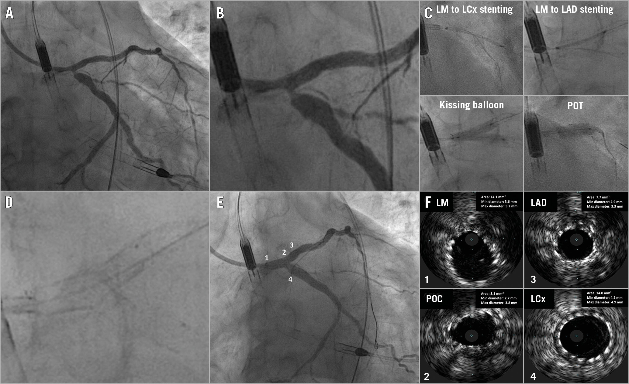
Figure 1. Culotte stenting technique of left main bifurcation with two self-apposing stents. A) & B) Coronary angiography. C) Left main (LM), left anterior descending (LAD), and LM-left circumflex (LCx) stenting: culotte technique, kissing balloon inflation and proximal optimisation technique (POT). D) & E) Angiographic result after stent deployment. F) Final intravascular ultrasound assessment confirming good stent apposition. POC: polygon of confluence
A 56-year-old male was admitted to our institution for percutaneous coronary intervention (PCI) of a left main (LM) bifurcation lesion. Since the LM dimensions were very large (reference vessel diameter [RVD] 5.1 mm on intravascular ultrasound [IVUS]), an aneurysm in the proximal left circumflex (LCx) (RVD 5.2 mm on IVUS) was present (Figure 1A, Figure 1B, Moving image 1) and no severe coronary calcification was evident, we chose to implant two self-apposing stents. Accordingly, after performing predilatation with semi-compliant balloons, a 3.5-4.5x22 mm sirolimus-eluting stent was deployed from the LM to the LCx and a 3.5-4.5x17 mm sirolimus-eluting stent was implanted from the LM to the left anterior descending (LAD) (using the culotte technique, Moving image 2, Moving image 3). After kissing balloon inflation and final proximal optimisation technique (Figure 1C, Moving image 4-Moving image 6), IVUS assessment confirmed good stent apposition on the LM, LAD and also on the proximal LCx aneurysm (Figure 1D-Figure 1F).
To the best of our knowledge, this is the first reported case showing the feasibility of two-stent LM bifurcation PCI using self-apposing stents. The self-expanding Xposition S sirolimus-eluting stent (Stentys, Paris, France) is mounted on a semi-compliant balloon, whose inflation splits the sheath in which the stent is restrained. The balloon and the sheath are then withdrawn leaving the nitinol stent apposed to the vessel wall. The advantages of this device when performing two-stent bifurcation PCI are the facilitated guidewire recrossing thanks to the open cell design, the cell disconnection feature allowing easier side branch access, and the possibility of achieving complete stent expansion and apposition, even in the presence of large vessel diameter discrepancies. Moreover, the highly visible markers at both stent edges allow accurate stent positioning, which is crucial when performing a two-stent strategy. On the other hand, the relatively large strut thickness (102 μm) could be a potential pitfall when performing a two-stent PCI resulting in a double strut layer. However, this is mitigated by large vessel dimensions and dynamic increase in stent diameter during the post-procedural period.
Conflict of interest statement
G. Tarantini reports honoraria for lectures/consulting from Medtronic, Edwards Lifesciences, Boston Scientific, GADA, Abbott. The other authors have no conflicts of interest to declare.
Supplementary data
To read the full content of this article, please download the PDF.
Moving image 1. Baseline coronary angiography.
Moving image 2. Sirolimus-eluting stent implantation from left main to left circumflex.
Moving image 3. Sirolimus-eluting stent implantation from left main to left anterior descending.
Moving image 4. Kissing balloon inflation.
Moving image 5. Final coronary angiography.
Moving image 6. Final coronary angiography.