A 55-year-old male underwent percutaneous coronary intervention (PCI) of a Medina 1,1,1 circumflex (LCx) and a first obtuse marginal (OM1) bifurcation lesion. A double kissing crush technique was performed, utilising a 2.75×12 mm everolimus-eluting stent (EES) in OM1 and a XIENCE V® 4.0×15 mm EES (Abbott Vascular, Santa Clara, CA, USA) in the LCx (Online Figure 1). Intravascular ultrasound (IVUS) and optical coherence tomography (OCT) runs were performed (Online Figure 2, Online Figure 3, Moving image 1). 3D reconstruction of the OCT further clarified the reconstruction of the bifurcation carina and paving, in particular in the crushed segment. Quantitative analysis showed only 10/327 struts to be malapposed (Figure 1, Moving image 2). We demonstrated the utility of OCT imaging in a complex bifurcation intervention.
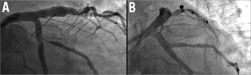
Figure 1. 3D reconstruction of the OCT run from the distal circumflex. A) An off-axis view aimed at the carina, revealing excellent reconstruction of the distal main vessel and side branch ostia; B) a view down the circumflex axis revealing a slight shift of the single-strut neocarina into the circumflex lumen; C) external view revealing optimal reconstruction of the side branch ostium.
Conflict of interest statement
V. Džavík has received unrestricted educational and research grants and speaker honoraria from Abbott Vascular. H.G. Bezerra has received speaker honoraria from St. Jude Medical. The other authors have no conflicts of interest to declare.
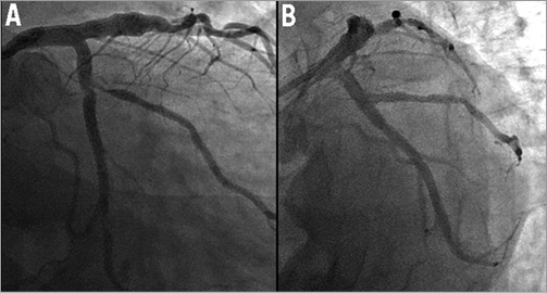
Online Figure 1. Pre- and post-PCI angiographic views of the LCx/OM1 bifurcation lesion. A) AP caudal view revealing a Medina 1,1,1 bifurcation lesion. B) Post bifurcation PCI image.
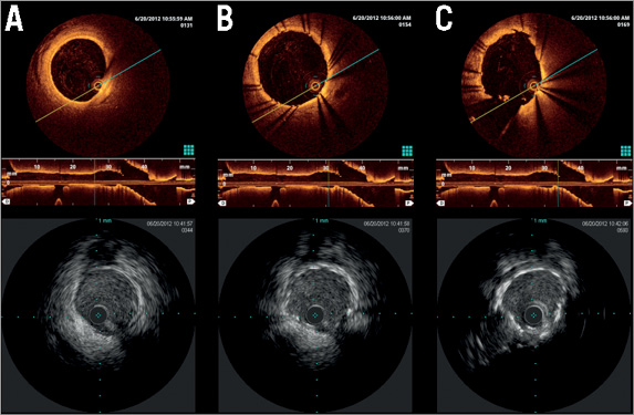
Online Figure 2. Post-PCI OCT and IVUS images in the circumflex, distal to the stent back to the bifurcation: A) mid circumflex distal to the stent; B) good stent strut apposition in the circumflex distal to the bifurcation; C) formation of a very short neocarina at the ostium of the OM1 with good stent strut apposition in the main vessel.
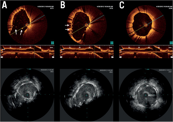
Online Figure 3. Post-PCI OCT and IVUS images of the circumflex from the bifurcation to the proximal aspect of the 4×15 mm stent. A) shows the formation of a short neocarina with three stent struts visible on OCT at 7 o’clock (arrow); B) a widely open OM1 ostium, “crushed” layers of stent struts visible at 9 o’clock on the OCT images (arrow); C) proximal aspect of the stent with optimal strut apposition.
Moving image 1. 2D OCT pullback through the circumflex artery.
Moving image 2. 3D flythrough from the proximal circumflex into the bifurcation.
Supplementary data
To read the full content of this article, please download the PDF.
Moving image 1. 2D OCT pullback through the circumflex artery.
Moving image 2. 3D flythrough from the proximal circumflex into the bifurcation.