IMAGE IN CARDIOLOGY

DOI: 10.4244/EIJY15M03_07

Interventional retrieval by catheter of a migrated atrial appendage occluder located in the arcus aortae

Nikola Bogunovic*, MSc; Siegfried Eckert, MD; Frank van Buuren, MD; Marios Vlachojannis, MD; Dieter Horstkotte, MD

We present the case of an 80-year-old male with atrial fibrillation with stroke/transient aphasia despite oral anticoagulation and an implanted Amplatzer LAA occluder (St. Jude Medical, St. Paul, MN, USA) from an external hospital. Control by transoesophageal echocardiography (TEE) before discharge revealed that the device had migrated into the aortic arch, almost completely obliterating the lumen of the aorta. The occluder’s orientation was lobe proximal, disc distal (Figure 1A-Figure 1C).

The aim was to capture the distally localised micro-screw of the disc. The disc maintains a distance between the lobe with its sharp stabilising wires at the edge and the aortic wall, as it has a greater diameter than the lobe.

The device (Figure 1D) was snared, but retraction into the sheath failed as the micro-screw of the device was blocking the manoeuvre (Figure 1E, Figure 1F). By retraction of the entire system into the abdominal aorta, the orientation of the device was modified, allowing it to be pulled back into the sheath (Figure 1G). On inspecting the retrieved occluder, no tissue was seen on the sharp wires (Moving image 1).

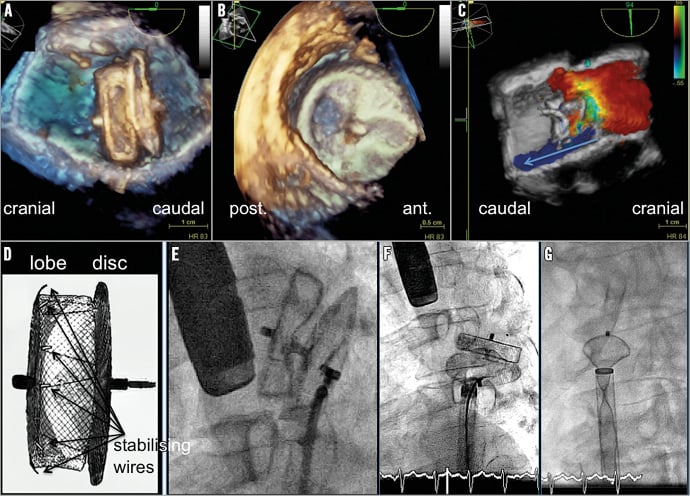
Figure 1. Multimodal imaging of the procedure. A-C) 3D transoesophageal echocardiogram (3D-TEE) demonstrating the occluder in the aortic long and short axis view. The occluder is localised within the descending part of the aortic arch. The diameter of the aorta is only 2 mm more than the width of the disc. The colour Doppler 3D-TEE demonstrates the reduced aortic flow passing the device. D) Construction of the Amplatzer Amulet left atrial appendage (LAA) occluder. The stabilising wires to fix the device within the LAA are marked (arrows). E) Fluoroscopic documentation of the device captured by a snare catheter at the micro-screw of the disc. F) The snare catheter gripped the micro-screw of the device from the side, which forced the device to move diagonally and therefore lateral to the entrance of the sheath. G) After further careful retrieval of the whole system (captured device, snare, sheath) into the abdominal aorta, the orientation of the device was optimised for retraction.

Conflict of interest statement

The authors have no conflicts of interest to declare.

Supplementary data

Moving image 1. Multimodal video sequence of the retrieval procedure.

Volume 11 Number 6
Oct 20, 2015
Volume 11 Number 6
View full issue


Key metrics

On the same subject

Short report

10.4244/EIJ-D-19-01088 Jun 11, 2021
Retrieval of embolised left atrial appendage closure devices
Kany S et al
free

CLINICAL RESEARCH

10.4244/EIJV11I13A292 Apr 20, 2016
Left atrial appendage occlusion with the AMPLATZER Amulet device: an expert consensus step-by-step approach
Tzikas A et al
free

Image – Interventional flashlight

10.4244/EIJ-D-21-00624 Jun 3, 2022
Percutaneous left atrial appendage closure in a surgically ligated left atrial appendage
Wong I et al
free

INTERVENTIONAL FLASHLIGHT

10.4244/EIJ-D-17-00871 Jan 19, 2018
Left atrial appendage closure: size does matter!
Achkouty G et al
free

IMAGE IN CARDIOLOGY

10.4244/EIJV12I2A44 Jun 10, 2016
The plug for the appendage
Buellesfeld L et al
free

10.4244/EIJV12SXA10 May 16, 2016
Left atrial appendage closure: patient, device and post-procedure drug selection
Tzikas A and Bergmann M
free

State-of-the-art

10.4244/EIJ-D-22-00627 Feb 6, 2023
Left atrial appendage occlusion
Holmes D et al
free
Trending articles
338.63

State-of-the-Art Review

10.4244/EIJ-D-21-00904 Apr 1, 2022
Antiplatelet therapy after percutaneous coronary intervention
Angiolillo D et al
free
295.45

Expert consensus

10.4244/EIJ-D-21-00898 Sep 20, 2022
Intravascular ultrasound guidance for lower extremity arterial and venous interventions
Secemsky E et al
free
226.03

State-of-the-Art Review

10.4244/EIJ-D-21-00426 Dec 3, 2021
Myocardial infarction with non-obstructive coronary artery disease
Lindahl B et al
free
209.5

State-of-the-Art Review

10.4244/EIJ-D-21-01034 Jun 3, 2022
Management of in-stent restenosis
Alfonso F et al
free
168.4

Expert review

10.4244/EIJ-D-21-00690 May 15, 2022
Crush techniques for percutaneous coronary intervention of bifurcation lesions
Moroni F et al
free
150.28

State-of-the-Art

10.4244/EIJ-D-22-00776 Apr 3, 2023
Computed tomographic angiography in coronary artery disease
Serruys PW et al
free
103.48

Expert consensus

10.4244/EIJ-E-22-00018 Dec 4, 2023
Definitions and Standardized Endpoints for Treatment of Coronary Bifurcations
Lunardi M et al
free
X

The Official Journal of EuroPCR and the European Association of Percutaneous Cardiovascular Interventions (EAPCI)

EuroPCR EAPCI
PCR ESC
Impact factor: 6.2
2022 Journal Citation Reports®
Science Edition (Clarivate Analytics, 2023)
Online ISSN 1969-6213 - Print ISSN 1774-024X
© 2005-2024 Europa Group - All rights reserved