
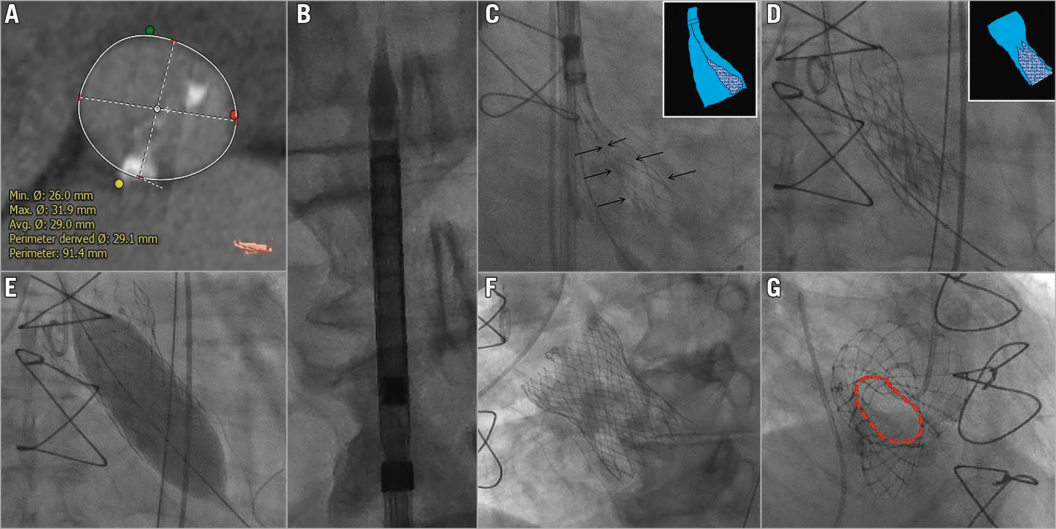
Figure 1. Computed tomography of native aortic annulus and serial fluoroscopic images of transcatheter aortic valve implantation procedure. A) Computed tomography of aortic annulus shows calcium at clock positions 2 and 6. B) In vitro fluoroscopic confirmation of the appropriately loaded 34 mm Evolut R valve system. C) Infolding of the valve frame is shown in between the arrows (dotted dark blue area in the cartoon inset). D) Underexpanded valve frame after complete release of the valve (dotted dark blue area in the cartoon inset). E) Balloon dilatation of the underexpanded valve frame with a 25 mm × 5 cm Z-MED II balloon. F) Complete expansion of the 34 mm Evolut R valve. G) The oval annulus of the completely expanded 34 mm Evolut R in en face view is shown as red dotted lines.
A 78-year-old gentleman had transcatheter aortic valve implantation using an Evolut™ R valve (Medtronic, Minneapolis, MN, USA). On computed tomography (CT) the perimeter-derived annulus diameter was 29.1 mm (Figure 1A). A 34 mm Evolut R was precisely loaded and the same was confirmed fluoroscopically before insertion into the sheath (Figure 1B). The Evolut R was recaptured twice due to unsatisfactory positioning. During the third release, following confirmation of annular position, the valve was deployed. Post deployment the patient had cardiac arrest and prompt cardiopulmonary resuscitation was initiated. It was then noticed that in the previous cine frame at 85% deployment there was infolding of the valve frame which had been overlooked (Figure 1C, Moving image 1). The underexpansion of the valve frame led to non-functioning of the Evolut R leaflets, which resulted in cardiac arrest (Figure 1D, Moving image 2). A swift post-dilatation with a 25 mm × 5 cm Z-MED™ II balloon (NuMED, Hopkinton, NY, USA) under rapid pacing expanded the valve completely with return of spontaneous circulation (Figure 1E, Figure 1F, Moving image 3). The en face view of the Evolut R valve post procedure confirmed the optimal expansion (Figure 1G, Moving image 4). In a previous report, infolding of the valve frame resulted in paravalvular leak but a normally functioning valve1. In a second report, infolding of the valve frame was recognised before complete release of the valve, hence it was recaptured and a new valve was implanted2. The nitinol memory of a large self-expanding valve (Evolut R; 34 mm) can be lost when there is severe annular calcium or prolonged exposure to higher core body temperature.
Conflict of interest statement
The authors have no conflicts of interest to declare.
Supplementary data
To read the full content of this article, please download the PDF.
Moving image 1. Infolding of the valve frame at 85% deployment.
Moving image 2. Underexpanded valve frame after complete release of the valve.
Moving image 3. Post-dilatation with a 25×50 mm Z-MED II balloon.
Moving image 4. En face view of the completely expanded valve.