Angiography before balloon pulmonary angioplasty (BPA) (Figure 1A, Figure 1C; Moving image 1, Moving image 2) detected a chronic total occlusion (CTO) lesion in the right pulmonary artery. Computed tomography (CT) images (Figure 1E, Figure 1G) implicated the distal vessel structures, suggesting that this CTO lesion existed just proximal to the bifurcation of three segmental branches. Furthermore, the stretched curved planar reformation (CPR) and straightened CPR images (Online Figure 1A, Online Figure 1C) demonstrated that two branches had a shared bifurcation point (yellow arrows), which could be distinguished from the ostium of another branch (red arrow). Wiring through the shared bifurcation point and the balloon dilation were performed. Angiography (Figure 1B, Figure 1D; Moving image 3, Moving image 4) and CT images (Figure 1F, Figure 1H, Online Figure 1B, Online Figure 1D) after BPA revealed revascularisation of the two branches (green arrowheads) and the remaining occlusion in another branch (blue arrowheads). Corresponding CT and CPR images are useful for identifying vessel structures distal to target lesions, positional relationship of a bifurcation and the ostia of branches, and estimated perfusion area. The CT-guided BPA procedure is therefore valuable for performing BPA safely and effectively.
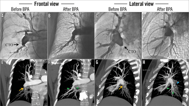
Figure 1. Revascularisation of chronic total occlusion lesion. Angiography (A & C) and corresponding computed tomography (CT) images (E & G) before balloon pulmonary angioplasty (BPA), and angiography (B & D) and CT images (F & H) after BPA.
Conflict of interest statement
The authors have no conflicts of interest to declare.
Supplementary data
Online Figure 1. Stretched curved planar reformation (CPR), before BPA (A) and after BPA (B), and straightened CPR images, before BPA (C) and after BPA (D). In panel D, the flows of the two treated segmental branches overlap.
Moving image 1. Frontal view of angiography before balloon pulmonary angioplasty (BPA).
Moving image 2. Lateral view of angiography before BPA.
Moving image 3. Frontal view of angiography after BPA.
Moving image 4. Lateral view of angiography after BPA.
Supplementary data
To read the full content of this article, please download the PDF.
Moving image 1. Frontal view of angiography before balloon pulmonary angioplasty (BPA).
Moving image 2. Lateral view of angiography before BPA.
Moving image 3. Frontal view of angiography after BPA.
Moving image 4. Lateral view of angiography after BPA.