Device description
Name and manufacturer: The HighLife transcatheter mitral valve replacement device; HighLife SAS, Paris, France.
Specific design: The HighLife transcatheter mitral valve replacement device is a two-component system. The valve prosthesis is implanted over a delivery system in the mitral position and is anchored by interacting and reaching an equilibrium position with a previously positioned subannular implant (SAI). The SAI is loose around the prosthesis; it blocks it from slipping into the atrium, whereas the inflow shape of the stent blocks it from slipping in the opposite direction into the ventricle. The sum of these opposite interactions results in a stable position in the heart (which could be described as an equilibrium position that adapts to different anatomies). The HighLife transcatheter mitral valve (TMV) is a stented mitral bioprosthesis made from glutaraldehyde cross-linked bovine pericardium mounted on a laser-cut nitinol frame. The SAI is intended to create a closed loop with a fixed perimeter around the patient’s native valve leaflets and chordae. In its final position, it allows the TMV to remain in a stable position in the heart, the native leaflets being positioned between the SAI and the TMV.
Procedural details
The current device is delivered via a transatrial or a transapical access (Figure 1). For both delivery routes, a first access is made to the femoral artery and an 18 Fr introducer sheath is positioned. This access enables the insertion of a specially designed catheter for the loop placement (LPC) and the delivery of the SAI. A second access is made either through an intercostal incision to the apex of the heart or via a right lateral minithoracotomy to the left atrium.
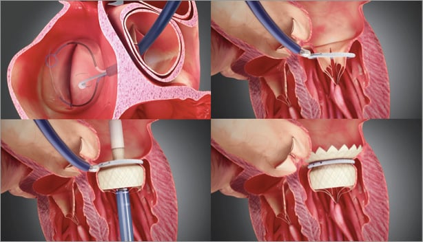
Figure 1. The LPC is advanced using a retrograde approach through the aortic valve. A guidewire loop is placed and both ends externalised. The delivery and closure of the SAI are carried out with a dedicated 18 Fr SAI delivery catheter (SDC) hosting the SAI and the two ends of the previously placed guidewire loop. The TMV delivery catheter is inserted through either the transapical (shown above) or the transatrial access. The outflow end of the TMV is deployed distal from the SAI and the SAI positioned close to the native annulus. Then, the inflow end of the TMV is deployed from the delivery catheter.
STEP 1. LOOP PLACEMENT
The LPC is advanced using a retrograde approach through the aortic valve inside the left ventricle. Two subannular tubes are deployed to reach around the subannular structure. A guidewire is caught with a snare, pulled back and externalised from the femoral access. As a result, a guidewire loop is placed, entering the femoral access travelling up to the left ventricle around the subannular apparatus and travelling back to the femoral access.
STEP 2. SAI DELIVERY
The delivery and closure of the SAI are carried out with a dedicated 18 Fr SAI delivery catheter (SDC) hosting the SAI and the two ends of the previously placed guidewire loop. The SDC is threaded over the two guidewire loop ends up to the left ventricle and the SAI is closed over the guidewire loop.
STEP 3. TMV DELIVERY
The TMV delivery catheter is inserted through either the transapical or the transatrial access. The catheter is advanced through the native mitral leaflets, and thus through the SAI positioned around the native leaflets. The outflow end of the TMV is deployed distal from the SAI. The catheter with the TMV attached is slightly pushed (transapical)/pulled (transatrial) towards the left atrium so as to position the SAI close to the native annulus. Then, the inflow end of the TMV is deployed from the delivery catheter. The catheter is closed and removed from the patient’s heart and the access to the apex/atrium is closed. The guidewire loop is removed from the SAI and the SAI closing catheter (SCC) by pulling on one end. The SCC is removed from the femoral access along with the introducer sheath and the femoral access is closed.
Animal studies
Acute and chronic animal studies have established the safety and feasibility of the procedure. In some chronic animals, the follow-up period was extended up to five months in order to evaluate the implants beyond the initial three-month period when anticoagulation was being administered (Figure 2). Histology showed good behaviour of the implants in vivo with adequate tissue ingrowth and limited mineralisation of the leaflets. When covered with a polyester sleeve, the SAI was completely encapsulated by ingrowing tissue, creating a strong natural seal and anchor. Absence of paravalvular regurgitation, left ventricular outflow tract obstruction and significant transvalvular gradients was found in the vast majority of animal experiments. Equivalence was shown between animals implanted over the transapical and transatrial access. Transseptal delivery was also successfully demonstrated with equivalent results.
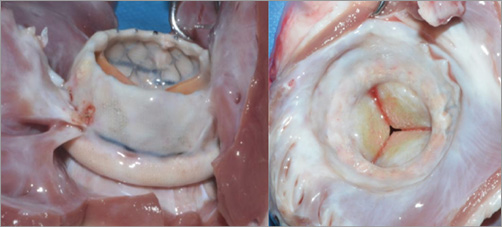
Figure 2. In chronic animal experiments up to five months, the HighLife TMV has shown complete encapsulation by ingrowing tissue creating a strong natural seal and anchor. A large inflow (“atrial” portion) helps to seal and prevent paravalvular mitral regurgitation. The outflow (“ventricular” portion) is short and designed to avoid left ventricular outflow tract obstruction.
Clinical data
The single-centre early feasibility clinical trial started in Kiev (Ukraine). A first patient was treated successfully and discharged home at day seven (Figure 3). Haemodynamics were satisfactory with no paravalvular leakage, trace central leak, 3 mmHg mean transvalvular gradient, an effective orifice area of 3.3 cm², and no gradient across the left ventricular outflow tract. The 30-day echocardiogram and 30-day CT scan demonstrated a properly functioning prosthesis in a stable position (Figure 4). Patient symptoms improved from Class III to Class I.
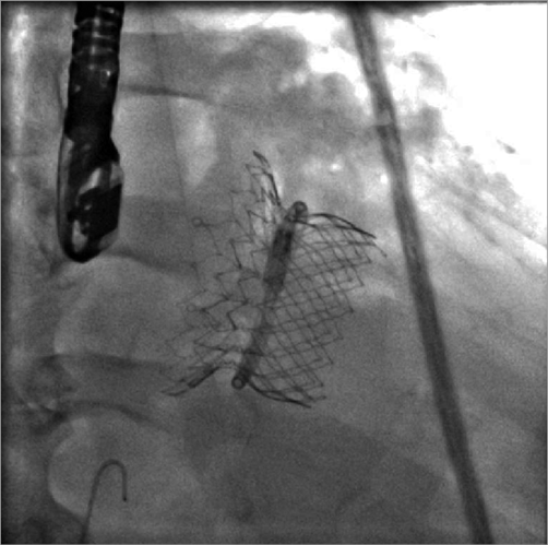
Figure 3. Patient 1. Acute fluoroscopic view of the valve and SAI in a stable position.
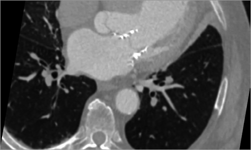
Figure 4. Patient 1. CT scan at 30 days shows good apposition of the inflow segment against the atrial wall, a patent LVOT and a stable position of the valve and SAI.
Unique features
The HighLife mitral transcatheter valve system consists of two components, a ring placed around the native leaflets (SAI) and a specifically designed stent with a groove placed inside the SAI. The SAI together with the native leaflets provides complete paravalvular sealing. Restrictive leaflet pathology is accommodated. Upon delivery, the valve stent centres itself within the SAI, facilitating potential transseptal delivery.
Potential improvements
A transseptal system is currently in development.
Conflict of interest statement
R. Lange is a consultant for and has an equity share in HighLife. N. Piazza is a consultant for and has an equity share in HighLife.
