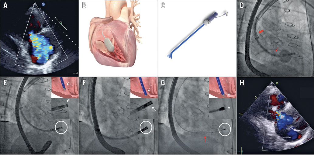

Figure 1. Novel features of the enhanced FORMA System. A) Baseline TTE showing massive TR. B) The FORMA System. C) & D) New steerable sheath (large arrow) facilitating delivery system advancement (small arrow) within the right ventricle. E) & F) Distal apposition indicator (white circle/arrow) inactivated (E) and activated (F) after contacting the myocardium. G) Final fluoroscopic position of the spacer (arrow), after anchor release (circle/asterisk). H) Post-procedural TTE demonstrating residual mild TR. TR: tricuspid regurgitation; TTE: transthoracic echocardiography
A 75-year-old man with a history of coronary bypass graft and aortic valve replacement presented with NYHA Class III dyspnoea and peripheral oedema despite diuretics. Echocardiography revealed massive functional tricuspid regurgitation (TR) due to moderate right ventricular (RV) and annular dilatation (Figure 1A), normally functioning aortic bioprosthesis and mild pulmonary hypertension. Following Heart Team evaluation (EuroSCORE II: 7%), compassionate transcatheter tricuspid repair with the enhanced FORMA System (Edwards Lifesciences, Irvine, CA, USA) was planned (Figure 1B).
Using general anaesthesia and left axillary vein puncture, the balloon-tipped delivery system was advanced through the 24 Fr steerable sheath (Figure 1C, Figure 1D). The anchor was deployed at the RV apex, under fluoroscopic visualisation of the apposition indicator (Figure 1E, Figure 1F), and an 18 mm spacer was positioned (Figure 1G) at the level of the tricuspid valve, reducing TR to mild (Figure 1H, Moving image 1-Moving image 5). The patient was discharged after 48 hours on anticoagulation and clinical improvement at 30 days allowed diuretic down-titration.
Despite satisfactory midterm results with the FORMA1, some anchoring system-related concerns have been raised. This case illustrates: (i) the 18 mm spacer covering large coaptation gaps and (ii) the main differentiating features of the enhanced FORMA System: (a) the new steerable guide sheath, for more coaxial alignment when dealing with large atrial cavities and (b) the redesigned delivery system with a radiopaque apposition indicator which enables tissue contact visualisation and may allow more predictable anchoring. Further studies need to explore whether such iterations may improve safety outcomes.
Conflict of interest statement
J. Rodés-Cabau has received institutional research grants from Edwards Lifesciences, and holds the Canadian Research Chair “Fondation Famille Jacques Larivière” for the Development of Structural Heart Disease Interventions. The other authors have no conflicts of interest to declare.
Supplementary data
To read the full content of this article, please download the PDF.
Moving image 1. Preprocedural TTE showing massive TR.
Moving image 2. Steering of the introducer sheath for coaxial delivery system alignment.
Moving image 3. Positioning of the delivery catheter (apposition indicator) and anchoring system release.
Moving image 4. Final positioning of the 18 mm spacer.
Moving image 5. Post-procedural TTE showing mild TR.

