
In 2013, a 78-year-old lady underwent surgery for reconstruction of her mitral valve with a 30 mm Carpentier-Edwards Physio annuloplasty ring (Edwards Lifesciences, Irvine, CA, USA) and reconstruction of her tricuspid valve with a 32 mm Carpentier-Edwards Physio tricuspid annuloplasty ring (Edwards Lifesciences). Gradually, a severe tricuspid regurgitation developed with heart failure of NYHA Class III.
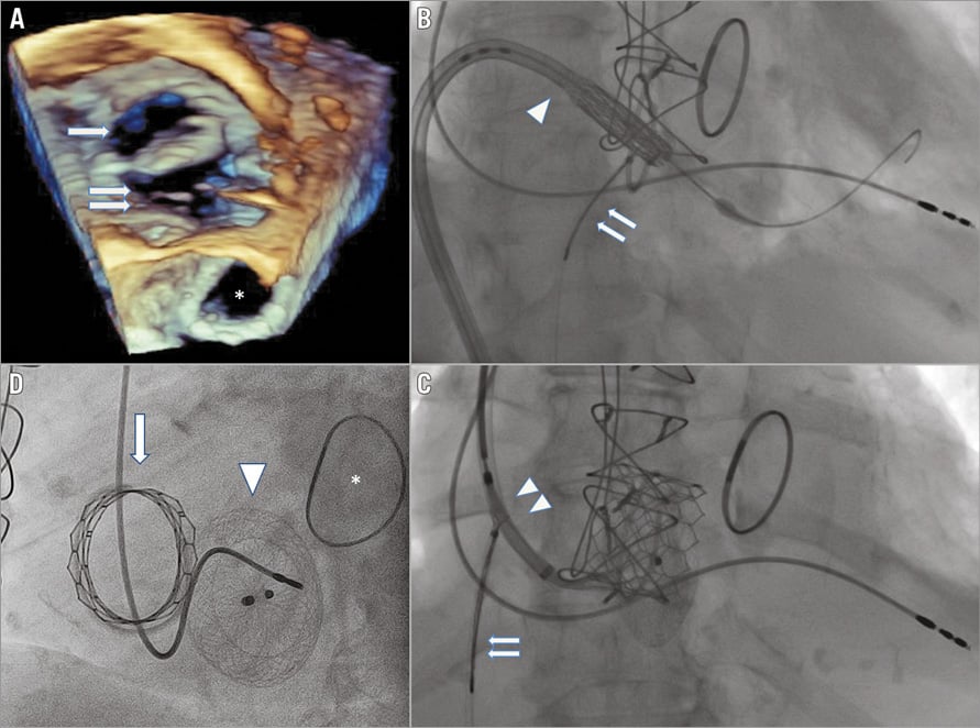
Current echocardiography demonstrated high-grade tricuspid regurgitation with both a valvular and a large paravalvular (PV) regurgitation (Panel A: arrow - tricuspid ring; double arrow - giant PV space sized 34×36 mm; *mitral ring). Considering the patient’s high risk, the Heart Team decided to attempt a percutaneous intervention.
Through a right femoral vein approach, a 29 mm Edwards SAPIEN 3 valve (Edwards Lifesciences) was introduced and positioned in the tricuspid valve ring. The help of a snare introduced through the contralateral femoral vein was required due to the difficult angle passing from the inferior vena cava (IVC) into the right ventricle (Panel B: arrowhead - SAPIEN 3; double arrow - snare).
For the next stage, we used a 28 mm AMPLATZER® Septal Occluder (St. Jude Medical, St. Paul, MN, USA) for sealing the paravalvular leak. Attempts to position the device through a right femoral vein approach failed; therefore, we switched to a right jugular vein approach to achieve a better angle. With the help of a snare introduced through the femoral vein (Panel C: double arrowhead - Occluder guide; double arrow – snare) (Moving image 1), the Occluder was placed and released in the correct position in the paravalvular space. Follow-up fluoroscopy showed the devices to be well positioned (Panel D: arrow - SAPIEN 3; arrowhead – Occluder; * mitral ring).
Percutaneous procedures of the tricuspid valve are difficult, mainly because of the angle of approach. The use of both the jugular and the femoral approach and the help of a snare may facilitate this procedure.
Conflict of interest statement
The authors have no conflicts of interest to declare.
Supplementary data
Moving image 1. Manipulation of the AMPLATZER Occluder delivery sheath introduced from the jugular approach with the help of a snare introduced from the femoral approach.
Supplementary data
To read the full content of this article, please download the PDF.
Moving image 1. Manipulation of the AMPLATZER Occluder delivery sheath introduced from the jugular approach with the help of a snare introduced from the femoral approach.

