DOI: 10.4244/EIJV9I12A249

Excimer laser for a highly stenotic saphenous vein graft: evidence of debulking by optical coherence tomography

Giampaolo Niccoli*, MD, PhD, FESC; Luca Di Vito, MD; Rocco Antonio Montone, MD; Italo Porto, MD, PhD; Filippo Crea, MD, PhD, FESC, FACC

A 73-year-old woman with a history of coronary artery bypass (saphenous vein graft [SVG] for left anterior descending artery [LAD] and right coronary artery) was admitted for non-ST-elevation acute coronary syndrome. Coronary angiography showed a proximal severe stenosis of the SVG for LAD that was the culprit lesion of the acute coronary syndrome (Figure 1A). Frequency-domain optical coherence tomography (FD-OCT) was performed using a non-occlusive technique and showed a highly fibrous stenosis of the SVG (MLA 0.7 mm2) (Figure 1B-E) that was debulked with excimer laser coronary atherectomy (ELCA) (Figure 1-A’,B’). However, a thrombus occurred at the lesion site immediately after ELCA (arrowhead), probably due to a dissection of the intimal layer (arrow) (Figure 1A’-E’,F,G,H). A drug-eluting stent was eventually implanted. After the procedure no rise in Troponin T levels was observed and she was asymptomatic at six months on double antiplatelet therapy.

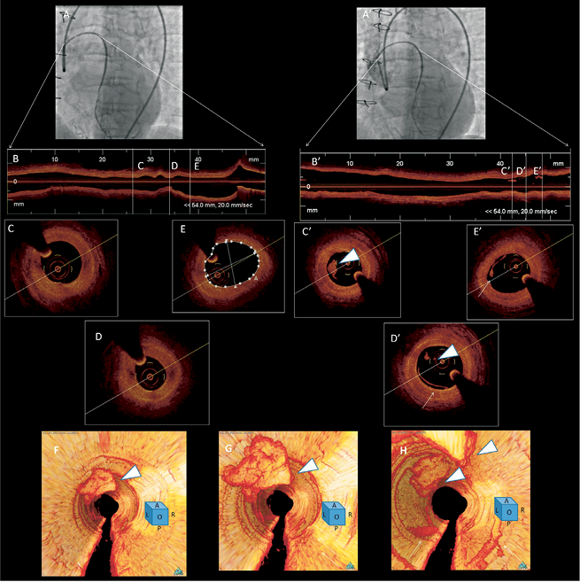
Figure 1. Angiographic and FD-OCT findings before and after ELCA (Vitesse 1.7; Spectranetics, Colorado Springs, CO, USA). FD-OCT was performed using a non-occlusive technique with 20 mm/sec pullback C7 XR machine (LightLab Imaging Inc./St Jude Medical, Westford, MA, USA).

SVG disease is associated with thrombi and cholesterol debris accumulation, and percutaneous interventional techniques in SVG are associated with significant risks of distal embolisation. This is, to our knowledge, the first report of intracoronary FD-OCT imaging showing debulking by ELCA of SVG lesions.

Conflict of interest statement

The authors have no conflicts of interest to declare.

Volume 9 Number 12
Apr 22, 2014
Volume 9 Number 12
View full issue


Key metrics

Suggested by Cory

10.4244/EIJV5I5A100 Nov 15, 2009
How should I treat a complex Post-CABG patient?
Sarno G et al
free

State-of-the-Art

10.4244/EIJ-D-21-00089 Jun 11, 2021
Intracoronary optical coherence tomography: state of the art and future directions
Ali ZA et al
free
Trending articles
202.45

State-of-the-Art

10.4244/EIJ-D-21-00089 Jun 11, 2021
Intracoronary optical coherence tomography: state of the art and future directions
Ali ZA et al
free
117

State-of-the-Art

10.4244/EIJ-D-24-00066 Apr 21, 2025
Management of complications after valvular interventions
Bansal A et al
free
71.8

State-of-the-art

10.4244/EIJ-D-22-00627 Feb 6, 2023
Left atrial appendage occlusion
Holmes D et al
free
68.9

State-of-the-Art

10.4244/EIJ-D-24-00992 Sep 15, 2025
Antithrombotic therapy in complex percutaneous coronary intervention
Castiello D et al
free
60.65

Clinical research

10.4244/EIJ-D-20-01155 Oct 20, 2021
A deep learning algorithm for detecting acute myocardial infarction
Liu W et al
free
59.45

Clinical research

10.4244/EIJ-D-23-00344 Sep 18, 2023
Clinical outcomes of TAVI with the Myval balloon-expandable valve for non-calcified aortic regurgitation
Sanchez-Luna JP et al
free
57.25

Expert Review

10.4244/EIJ-D-25-00201 Oct 10, 2025
Drug-coated balloons for coronary bifurcation lesions
Fezzi S et al
free
57.25

Expert Review

10.4244/EIJ-D-25-00201 Oct 20, 2025
Drug-coated balloons for coronary bifurcation lesions
Fezzi S et al
free
49.55

CLINICAL RESEARCH

10.4244/EIJ-D-17-00962 Apr 6, 2018
A new optical coherence tomography-based calcium scoring system to predict stent underexpansion
Fujino A et al
free
X

PCR
Impact factor: 9.5
2024 Journal Citation Reports®
Science Edition (Clarivate Analytics, 2025)
Online ISSN 1969-6213 - Print ISSN 1774-024X
© 2005-2025 Europa Group - All rights reserved