IMAGE IN CARDIOLOGY

DOI: 10.4244/EIJV10I12A253

Coronary perforation after bioresorbable vascular scaffold implantation

Josep Gomez-Lara*, MD, PhD; Luis Teruel, MD; Rafael Romaguera, MD; Gerard Roura, MD; José-Luis Ferreiro, MD; Guillermo Sanchez-Elvira, MD; Joan-Antoni Gomez-Hospital, MD, PhD; Angel Cequier, MD, PhD

A 67-year-old-man with hypertension and hypercholesterolaemia presented with unstable angina. The patient was admitted and studied with exercise echocardiography showing normal left ventricular function and moderate hypokinesia in the posterior wall during exercise. The coronarography showed a severe lesion in the right posterior descending coronary artery.

The patient was informed of the ABSORB II trial and agreed to participate in the study. The QCA-derived proximal and distal maximal luminal diameters of the segment to be scaffolded were 2.79 and 2.87 mm, respectively. After predilatation with a 2.5×14 mm semi-compliant balloon at low pressure, ECG-gated 45 MHz IVUS imaging was performed (Revolution®; Volcano Corp., Rancho Cordova, CA, USA). The greyscale and virtual histology images showed a severely fibro-calcified lesion with important negative remodelling (35% with respect to the proximal and distal healthy regions).

After IVUS imaging, a new predilatation was performed showing appropriate balloon expansion at 8 atm. Then, an Absorb® 3.0×18 mm BVS (Abbott Vascular, Santa Clara, CA, USA) was implanted (14 atm). Immediately after implantation the patient had severe chest pain. The angiography showed an Ellis type III perforation within the scaffolded segment at the site with negative remodelling. Prolonged balloon inflation and reversion of the anticoagulation with protamine was performed. After five minutes, the perforation was sealed and no further interventions were required. The patient indicated no chest pain and the artery had TIMI 3 flow.

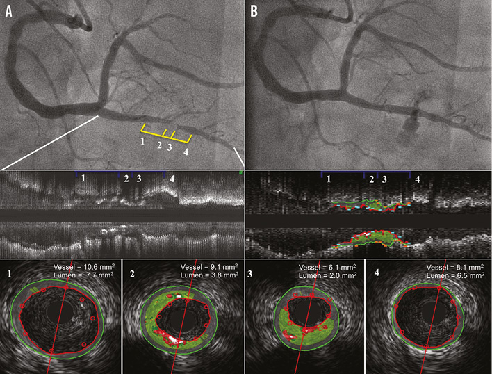
Figure 1. Angiographic and intravascular ultrasound images prior to and after scaffold implantation. A) Severe lesion in the posterior descending coronary artery. B) Coronary perforation immediately after Absorb scaffold implantation. 1) to 4) IVUS images of the posterior descending coronary artery lesion after predilatation.

Conflict of interest statement

The authors have no conflicts of interest to declare.

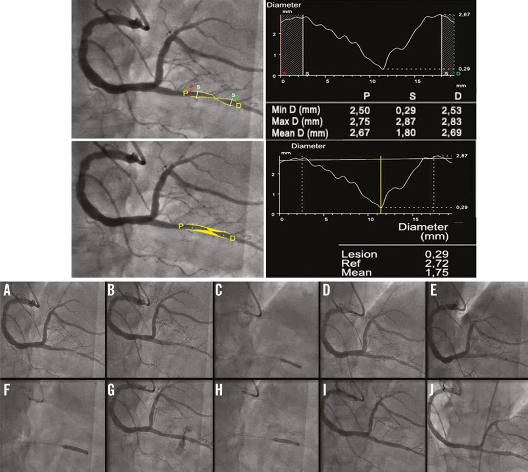
Appendix Figure 1. QCA measurements before scaffold implantation (top) and angiographic images of the coronary intervention and one-month follow-up control (bottom). A) Atherosclerotic lesion located in posterior descending coronary artery. B) Positioning of a compliant balloon 2.5×14 mm. C) Balloon inflation at 8 atm. D) Angiographic result after balloon predilatation. E) Positioning of a 3.0×18 mm bioresorbable scaffold. F) Scaffold implantation at 14 atm. G) Coronary perforation within the scaffolded segment. H) Prolonged balloon inflation (4 mins) and reversion of the anticoagulation with protamine. I) Final angiographic result at index procedure. J) Angiographic control at one-month follow-up.

Volume 10 Number 12
Apr 20, 2015
Volume 10 Number 12
View full issue


Key metrics

Suggested by Cory

IMAGE IN CARDIOLOGY

10.4244/EIJV12I1A10 May 16, 2016
Coronary aneurysm without malapposition after bioresorbable vascular scaffold implantation
Timmers L et al
free

IMAGE IN CARDIOLOGY

10.4244/EIJV10I11A225 Mar 20, 2015
The “plastic healing concept”: implantation of bioabsorbable scaffolds in spontaneous coronary artery dissection
Al Mamary A et al
free

CLINICAL RESEARCH

10.4244/EIJ-D-17-00246 Jul 20, 2017
One-year clinical results of the Italian diffuse/multivessel disease ABSORB prospective registry (IT-DISAPPEARS)
Testa L et al
free

10.4244/EIJV11SVA42 May 19, 2015
Treatment of bioresorbable scaffold failure
Felix C et al
free

CLINICAL RESEARCH

10.4244/EIJV10I4A78 Aug 19, 2014
Lessons learned from acute and late scaffold failures in the ABSORB EXTEND trial
Ishibashi Y et al
free

IMAGE IN CARDIOLOGY

10.4244/EIJV11I1A14 May 19, 2015
Very late bioresorbable vascular scaffold thrombosis: a new clinical entity
Azzalini L et al
free

IMAGE IN CARDIOLOGY

10.4244/EIJ-D-16-00259 Mar 20, 2017
Neoatherosclerosis: an emerging and conceptually unexpected cause of very late bioresorbable vascular scaffold failure
Hiltrop N et al
free
Trending articles
318.1

State-of-the-Art Review

10.4244/EIJ-D-21-00695 Nov 19, 2021
Transcatheter treatment for tricuspid valve disease
Praz F et al
free
116.75

State-of-the-Art

10.4244/EIJ-D-24-00066 Apr 21, 2025
Management of complications after valvular interventions
Bansal A et al
free
108.3

Viewpoint

10.4244/EIJ-E-22-00007 May 15, 2022
TAVI at 20: how a crazy idea led to a clinical revolution
Eltchaninoff H et al
free
91.6

Image – Interventional flashlight

10.4244/EIJ-D-22-00344 Aug 5, 2022
First dedicated transcatheter leaflet splitting device: the ShortCut device
Tchétché D et al
free
71.3

State-of-the-art

10.4244/EIJ-D-22-00627 Feb 6, 2023
Left atrial appendage occlusion
Holmes D et al
free
68.9

State-of-the-Art

10.4244/EIJ-D-24-00992 Sep 15, 2025
Antithrombotic therapy in complex percutaneous coronary intervention
Castiello D et al
free
60.65

Clinical research

10.4244/EIJ-D-20-01155 Oct 20, 2021
A deep learning algorithm for detecting acute myocardial infarction
Liu W et al
free
49.55

CLINICAL RESEARCH

10.4244/EIJ-D-17-00962 Apr 6, 2018
A new optical coherence tomography-based calcium scoring system to predict stent underexpansion
Fujino A et al
free
43.45

State-of-the-Art Review

10.4244/EIJ-D-21-00145 Sep 20, 2021
Robotics, imaging, and artificial intelligence in the catheterisation laboratory
Beyar R et al
free
X

PCR
Impact factor: 9.5
2024 Journal Citation Reports®
Science Edition (Clarivate Analytics, 2025)
Online ISSN 1969-6213 - Print ISSN 1774-024X
© 2005-2025 Europa Group - All rights reserved