DOI: 10.4244/EIJV7I8A158

A drug-eluting stent, accidentally removed as late as 1½ years after implantation

Wim J. van der Giessen†1,2*, MD, PhD; Gökhan Ertaş1, MD; Heleen M.M. van Beusekom1, PhD; Carl Schultz1, MD; Jurgen Ligthart1, BSc; Marc van der Linden1, MD, PhD; Evelyn Regar1, MD, PhD

A 71-year-old male patient underwent PCI for an occluded everolimus-eluting stent implanted 17 months earlier (Figure top panel A). Following balloon angioplasty, directional atherectomy was performed to aid in-stent placement. After five cuts the atherectomy catheter could only be withdrawn with considerable resistance, retrieving a deformed stent containing neointima (Figure bottom panel). Angiography confirmed the absence of the stent and a patent LAD without contrast extravasation, but with hazy borders (Figure top panel B). Endovascular imaging showed a dissected LAD with intraluminal flaps (Figure middle panels A-D) that were successfully stented (Figure top panel C). The patient showed no symptoms or biomarker release during and after the procedure. Histology of stent and atherectomy tissue showed an inhomogeneous distribution of heavily vascularised myxoid and fibrous tissue containing both old (hemosiderin) and recent (erythrocyte ghosts) neointimal bleeding. Six month follow-up angiography showed a nice result (Figure top panel D).
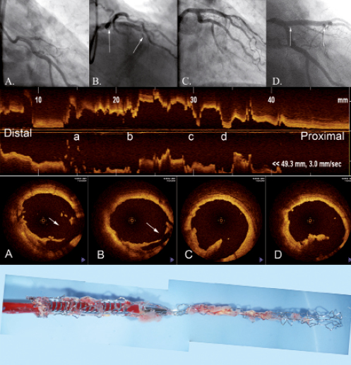
Figure 1. Top panel: angiogram of procedure; middle panel: OCT imaging; bottom panel: deformed stent containing neointima.

Conclusion

Atherectomy can be useful to reduce in-stent neointima burden prior to stenting for in-stent restenosis.1 Stent removal by atherectomy is a rare complication.2 The present case illustrates substantial intimal damage but without transmural vascular disruption.

Conflict of interest statement

The authors have no conflict of interest to declare.
Volume 7 Number 8
Dec 30, 2011
Volume 7 Number 8
View full issue


Key metrics

Suggested by Cory

10.4244/EIJV7I8A141 Dec 30, 2011
Complications with a happy ending
Muller O et al
free

10.4244/EIJV7I3A65 Jul 29, 2011
How should I treatsevere calcific coronary artery disease?
Fernandez J et al
free

IMAGE IN CARDIOLOGY

10.4244/EIJV12I10A201 Nov 20, 2016
Ruptured “non-culprit” in-stent neoatherosclerosis during ST-segment elevation acute myocardial infarction
Cuesta J et al
free

Image – Interventional flashlight

10.4244/EIJ-D-20-00507 Aug 6, 2021
Optical coherence tomography and tailored treatment of in-stent restenosis
Jia H et al
free

Clinical research

10.4244/EIJ-D-20-00539 Aug 27, 2021
Predictors and outcomes of neoatherosclerosis in patients with in-stent restenosis
Nakamura D et al
free
Trending articles
151.43

State-of-the-Art

10.4244/EIJ-D-22-00776 Apr 3, 2023
Computed tomographic angiography in coronary artery disease
Serruys PW et al
free
55.9

Clinical research

10.4244/EIJ-D-22-00621 Feb 20, 2023
Long-term changes in coronary physiology after aortic valve replacement
Sabbah M et al
free
54.9

Expert review

10.4244/EIJ-D-21-01010 Jun 24, 2022
Device-related thrombus following left atrial appendage occlusion
Simard T et al
free
43.75

Clinical Research

10.4244/EIJ-D-21-01091 Aug 5, 2022
Lifetime management of patients with symptomatic severe aortic stenosis: a computed tomography simulation study
Medranda G et al
free
39.95

Clinical research

10.4244/EIJ-D-22-00558 Feb 6, 2023
Permanent pacemaker implantation and left bundle branch block with self-expanding valves – a SCOPE 2 subanalysis
Pellegrini C et al
free
X

The Official Journal of EuroPCR and the European Association of Percutaneous Cardiovascular Interventions (EAPCI)

EuroPCR EAPCI
PCR ESC
Impact factor: 7.6
2023 Journal Citation Reports®
Science Edition (Clarivate Analytics, 2024)
Online ISSN 1969-6213 - Print ISSN 1774-024X
© 2005-2024 Europa Group - All rights reserved