DOI:

Slow flow after stenting of a coronary lesion with a large lipid core plaque detected by near-infrared spectroscopy

Bilal Saeed1, MD; Subhash Banerjee2, MD, FSCAI; Emmanouil S. Brilakis2*, MD, PhD, FSCAI

Coronary angiography of a patient with non-ST segment elevation acute myocardial infarction, demonstrating an eccentric mid left anterior descending artery lesion (Panel A) associated with a large lipid core plaque by near-infrared spectroscopy (Panel B) and severe luminal stenosis by intravascular ultrasonography (Panel C). The results of near-infrared spectroscopy are presented as a chemogram, where yellow indicates lipid core plaque at longitudinal (x-axis) and rotational (y-axis) locations. After the lesion was stented with a 3.5 x 28 mm everolimus-eluting stent (Panel D), the patient developed slow flow and subsequent NIRS showed that the size of the lipid core had decreased (Panel E). IVUS revealed an intra-stent luminal filling defect suggesting massive plaque prolapse (arrows in Panel F), associated with a strong signal of lipid core by NIRS. Intracoronary nicardipine, and several attempts with aspiration and rheolytic thrombectomy failed to improve the flow and the filling defect persisted. The patient’s troponin T increased from 0.3 ng/mL before stenting to 0.7 ng/mL the day following intervention. The findings strongly suggest that the mechanism for slow flow in this case might have been distal embolisation of the lipidic content of the plaque.

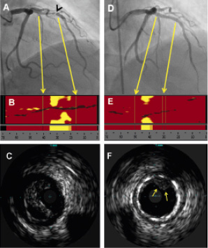
Figure 1. Angiographic, near-infrared spectroscopy, and intravascular ultrasonography images from a patient who developed slow flow during percutaneous coronary intervention.

Volume 6 Number 4
Sep 30, 2010
Volume 6 Number 4
View full issue


Key metrics

Suggested by Cory

IMAGE IN CARDIOLOGY

10.4244/EIJV11I7A157 Nov 20, 2015
Stent failure due to simultaneous aggressive neoatherosclerosis of first- and current-generation drug-eluting stents
Komukai K et al
free

IMAGE IN CARDIOLOGY

10.4244/EIJV12I10A201 Nov 20, 2016
Ruptured “non-culprit” in-stent neoatherosclerosis during ST-segment elevation acute myocardial infarction
Cuesta J et al
free

Clinical research

10.4244/EIJ-D-20-01421 Dec 17, 2021
Near-infrared spectroscopy to predict microvascular obstruction after primary percutaneous coronary intervention
Terada K et al
free
Trending articles
310.93

State-of-the-Art Review

10.4244/EIJ-D-21-00695 Nov 19, 2021
Transcatheter treatment for tricuspid valve disease
Praz F et al
free
92.2

State-of-the-Art Review

10.4244/EIJ-D-20-01296 Aug 27, 2021
Management of cardiogenic shock
Thiele H et al
free
76.25

State-of-the-Art

10.4244/EIJ-D-23-00840 Sep 2, 2024
Aortic regurgitation: from mechanisms to management
Baumbach A et al
free
56.65

Clinical research

10.4244/EIJ-D-20-01155 Oct 20, 2021
A deep learning algorithm for detecting acute myocardial infarction
Liu W et al
free
35

Original Research

10.4244/EIJ-D-25-00331 May 21, 2025
One-month dual antiplatelet therapy followed by prasugrel monotherapy at a reduced dose: the 4D-ACS randomised trial
Jang Y et al
open access
26

Expert Review

10.4244/EIJ-D-24-00535 May 5, 2025
Catheter-based techniques for pulmonary embolism treatment
Costa F et al
X

The Official Journal of EuroPCR and the European Association of Percutaneous Cardiovascular Interventions (EAPCI)

EuroPCR EAPCI
PCR ESC
Impact factor: 9.5
2024 Journal Citation Reports®
Science Edition (Clarivate Analytics, 2025)
Online ISSN 1969-6213 - Print ISSN 1774-024X
© 2005-2025 Europa Group - All rights reserved